
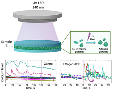
 Top: Schematic model of experimental setup for optical platelet activation. Bottom left: Control experiments without caged ADP, showing that two UV flashes don’t influence platelet calcium signaling. Bottom right: Several typical signals obtained during experiments with caged ADP.
Blood platelets are cells that provide hemostasis, stopping blood flow and thus ensuring that blood will not leak through any small damage of the blood vessel that might occur. The hemostatic reaction starts with platelet activation, triggered when the platelets sense stimuli endemic to the environment outside of the blood vessel. The study of platelet activation is important, as this process plays a significant role in many pathological conditions. In work published this year, we introduced an optical method that can provide a superior insight into the dynamics of platelet activation.
The hallmark of platelet activation is a spiking of calcium ion concentrations in the cytoplasm. This pattern allows one to trace the dynamics of platelet activation using real-time calcium probes. The activation itself can be triggered in vitro by the addition of adenosine diphosphate (ADP), a substance that is liberated from damaged cells during vessel wall injury, as platelets have special receptors for ADP. This method, introduced in pioneering work,1 led to a quantitative understanding of the platelet activation process.2,3 However, in these studies platelets were immobilized onto a surface, which alters the properties of cells and hence the dynamics of activation.
Recently, we have overcome this deficiency through an experimental protocol in which platelet activation is triggered by an optical pulse.4 Our method enables the study of activation without attachment of platelets to a surface, because there is no displacement caused by the addition of an agonist. The protocol is based on ADP that is “caged” by a photoactive compound and released when illuminated by UV light. The system allows one to track the very early stages of platelet activation in single, free-moving cells.
In our approach, the caged ADP is mixed with a suspension of platelets before the experiment. It doesn’t bind to the ADP receptors until a UV flash is applied, allowing diffusion and binding to the ADP receptor to be separated in time. We performed UV illumination using a 340-nm, 3-W LED connected to an Arduino board to allow control of the flash duration.
Our experiments have demonstrated, for the first time to our knowledge, accurate single-cell measurements of platelet activation in its early phase. The described approach opens new avenues in platelet research, including the study of intercellular signal transmission and nonlinear interaction of different biochemical pathways. We believe that the approach we have described will advance the study of platelet function in various physiological and pathological conditions, and help in the diagnosis of cardiovascular diseases.
The authors acknowledge support from RussianScience Foundation, grant #18-15-00049.
Researchers
Darya V. Spiryova, Alexei Yu. Vorobev, Vadim V. Klimontov, Elena A. Koroleva and Alexander E. Moskalensky, Novosibirsk State University, Novosibirsk, Russia
References
1. J.W. Heemskerk et. al. Biochem. J. 283, 379 (1992).
2. J.E. Purvis et al. Blood 112, 4069 (2008).
3. A.N. Sveshnikova et. al. Mol. Biosyst. 11, 1052 (2015).
4. D.V. Spiryova et al. Biomed. Opt. Express 11, 3319 (2020).
