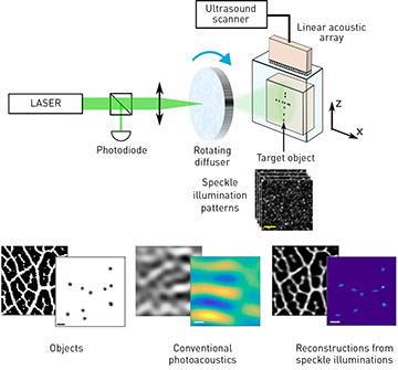
 Top: Experimental setup. A pulsed laser beam produces temporally varying, unknown random speckle patterns that illuminate the target; for each speckle pattern, photoacoustically generated ultrasound waves from the sample’s absorbing portions are recorded by a linear transducer array. Bottom: Results comparing the target objects (left), the images obtained with conventional photoacoustics (center), and the images obtained using our super-resolution approach (right). Background, numerical results; foreground, experimental results.
Optical microscopy, perhaps the most important tool in biomedical investigation, is currently limited to imaging depths of a few hundred microns inside tissue due to light scattering. All techniques that can go beyond these depths suffer from inferior resolution. Of these, a leading approach for deep-tissue optical imaging is photoacoustic imaging, in which ultrasonic waves that are produced as a result of light absorption are used to form images of deep-lying structures. At depths beyond one millimeter, however, acoustic diffraction limits the resolution of photoacoustic imaging to, at best, an order of magnitude poorer than the optical diffraction resolution limit.
In a recent work, we have shown that the conventional acoustic diffraction limit can be surpassed by combining dynamic optical speckle illumination with advanced computational reconstruction algorithms based on compressed sensing.1 Our approach’s improved resolution originates from two sources. The first is the uncorrelated photoacoustic signal fluctuations induced by the dynamic speckle illumination.2 These are analogous to the fluorescence fluctuations used for super-resolution optical fluctuation imaging (SOFI) in fluorescence microscopy.3
The second source for super-resolution arises from a compressed-sensing reconstruction framework. We have shown that exploiting inherent priors on the (unknown) speckle illumination patterns and the imaged object—such as their non-negativity and structural sparsity—makes it possible to obtain a substantial increase in imaging fidelity and a considerable reduction in acquisition time, beyond what is possible with speckle fluctuations alone.1 Our results may open the path to previously impossible optical investigations of deep-lying structures.
Researchers
E. Hojman and O. Katz, The Hebrew University of Jerusalem, Israel
T. Chaigne, Charité Universitätsmedizin Berlin and Humboldt University, Einstein Center for Neuroscience, NeuroCure Cluster of Excellence,
Berlin, Germany
S. Gigan, Laboratoire Kastler-Brossel, Université Pierre et Marie Curie, Paris, France
O. Solomon and Y.C. Eldar, Technion, Haifa, Israel
E. Bossy, University Grenoble Alpes, CNRS, LIPhy, Grenoble, France
References
1. E. Hojman et al. Opt. Express 25, 4875 (2017).
2. T. Chaigne et al. Optica 3, 54 (2016).
3. T. Dertinger et al. Proc. Natl. Acad. Sci. USA 106, 22287 (2009).
