
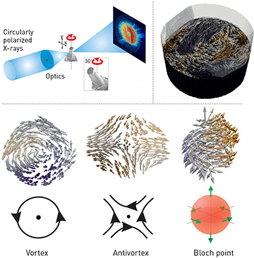
 Top: Experimental setup for magnetic tomography (left), and reconstructed internal magnetic structure of the pillar (right). Bottom: topological magnetic structures observed in the pillar, including experimental reconstruction and accompanying schematic.
Determining the internal 3-D configuration of magnetization and magnetic nanotextures is important not only for understanding magnetism, but also for the improved performance of magnets in applications such as motors, energy harvesting and data storage. Magnetic tomography using neutron magnetic imaging can map the internal magnetic structure within millimeter-sized magnets at a spatial resolution of tens of microns,1 and soft-X-ray2 and electron magnetic tomography have achieved nanoscale imaging of micromagnetic details within thin films and nanostructures. Until now, however, mapping the 3-D internal magnetization vector field at the nanoscale within magnets thicker than a few hundred nanometers has not been possible.
We recently demonstrated hard-X-ray magnetic tomography, a technique that applies the advantages of hard X-rays for tomographic imaging to magnetic systems, and that makes it possible to determine internal magnetic structures in 3-D. In particular, higher-energy, hard-X-ray photons offer both a high penetration depth and a high spatial resolution that are ideal for investigating such structures at the nanoscale, and that have enabled 14.6-nm spatial resolution in non-magnetic investigations.3
We had previously demonstrated hard-X-ray magnetic imaging at the nanoscale using resonant circularly polarized X-rays to probe the X-ray magnetic circular dichroism of a sample, and thus the single component of the magnetization parallel to the X-ray beam.4 In our most recent work, we performed dual-rotation-axis tomography, providing sensitivity to all three vector components of the magnetization.5
Reconstructing the internal magnetization constitutes a significant challenge, as magnetic tomography (unlike traditional tomography) requires retrieving three components of magnetization for each voxel. Previous studies incorporated some prior knowledge of the sample’s magnetic properties.2 In our more recent work, we developed an iterative reconstruction algorithm that does not require any assumptions about the system’s magnetic properties.
Using these principles, we reconstructed the internal magnetization configuration of a magnetic GdCo2 micropillar of diameter 5 µm, at a spatial resolution of 100 nm, using hard-X-ray magnetic tomography. Within the pillar, we observed smoothly varying 3-D magnetic patterns, containing a variety of fundamental topological structures such as vortices, antivortices and domain walls, which intersect to form a complex magnetic configuration. At the intersections of these structures, we also found magnetization singularities, or Bloch points. These points were predicted theoretically more than 50 years ago, but their surrounding structure has not been observed directly until now.
These techniques should allow investigation of even smaller magnetization details with the development of next-generation synchrotrons, which could allow spatial resolutions that are up to five times higher.
Researchers
Claire Donnelly, Valerio Scagnoli and Laura J. Heyderman, ETH Zurich and Paul Scherrer Institute, Switzerland
Manuel Guizar-Sicairos, Mirko Holler and Jörg Raabe, Paul Scherrer Institute
Sebastian Gliga, University of Glasgow, U.K.
References
1. I. Manke et al. Nat. Commun. 1, 125 (2010).
2. R. Streubel et al. Nat. Commun. 6, 7612 (2015).
3. M. Holler et al. Nature 543, 402 (2017).
4. C. Donnelly et al. Phys. Rev. B 94, 064421 (2016).
5. C. Donnelly et al. Nature 547, 328 (2017).
