
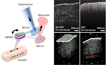
 Left: Imaging scheme of DA-OCT system. Top right: Representative images of porcine ear skin acquired by DA-OCT and conventional OCT. E, epidermis; D, dermis. Bottom right: DA-OCT 3-D volume snapshots of porcine ear skin sample. V, blood vessel; S, subcutaneous tissue. (See online video for full 3-D volume of the sample.)
Light scattering in tissue fundamentally limits the penetration depth of almost all optical imaging techniques. Many conventional imaging methods rely on separating the component of returned light that has been scattered a single time. However, these methods struggle to provide satisfying images beyond the most superficial layers. Interferometric imaging techniques, such as optical coherence tomography (OCT), offer imaging at extended depths using the coherence properties of light. Yet depth penetration in soft tissues is still typically below 1 mm, and full-thickness imaging of human skin is not possible.
We have recently developed dual-axis optical coherence tomography (DA-OCT) for deep-tissue imaging.1 The DA-OCT system has a center wavelength of 770 nm and a bandwidth of 108 nm, and uses an off-axis illumination/detection configuration to preferentially detect multiply forward-scattered photons from deep tissue.2,3 In our recent work, a microelectromechanical-system (MEMS) mirror laterally scans the beam across the sample while maintaining the off-axis illumination/detection geometry. This allows a fast acquisition speed (56 μs line time) that opens the door to both in vivo and volumetric imaging capabilities.1
Imaging of porcine ear skin tissue ex vivo shows DA-OCT’s increased penetration depth. We acquired a 512×300×2048-voxel (1.2×1.2×2-mm) image volume in less than 4 minutes. This depth improvement by DA-OCT allows visualization of deep-tissue structures otherwise inaccessible by conventional OCT imaging. Dermal structures (epidermis and dermis) are clearly resolved, and subsurface features such as blood vessels and subcutaneous tissue can also be identified.
DA-OCT offers the possibility of effective in vivo imaging of deep tissues. Although the technique sacrifices some axial resolution by the detection of multiply forward-scattered photons, we have demonstrated that it can provide deep-tissue-imaging capabilities significantly beyond those offered by conventional OCT systems. Spectroscopic contrast can also provide greater diagnostic capabilities for various clinical applications in surgery and dermatology, while imaging in other spectral windows, such as near 1300 nm, can further extend penetration depths.
Researchers
Yang Zhao and Adam Wax, Duke University, N.C., USA
References
1. Y. Zhao et al. Opt. Lett. 42, 2302 (2017).
2. T.E. Matthews et al. Optica 1, 105 (2014).
3. T.E. Matthews et al. Appl. Opt. 52, 8220 (2013).
