

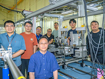
The research team that developed ultra-low-light megapixel X-ray ghost imaging, led by Tiqiao Xiao. [Image: Tiqiao Xiao, Shanghai Advanced Research Institute, Chinese Academy of Sciences]
Many patients wonder if the X-rays ordered by their physicians will subject them to radiation overdoses. In the future, those medical images could require a lot less radiation.
Scientists in China have developed a new computational imaging technique that reconstructs X-ray pictures from a tiny fraction of the photons normally required in medical diagnostics. (Optica, doi: 10.1364/OPTICA.598975). The proof-of-concept study yielded images nearly 2 megapixels in resolution, yet using only 0.48% of the usual X-ray dosage.
“While traditional X-ray imaging relies on enough X-ray photons reaching a detector to form a clear image, our approach uses computational techniques to reconstruct an image from fewer photons,” said researcher Tiqiao Xiao, Shanghai Advanced Research Institute, Chinese Academy of Sciences. “We were able to show the low-dose potential of this approach by achieving megapixel radiology with ultra-low-light.”
The research team hopes that the technology could make future medical X-rays safer for children and pregnant patients, plus anyone who must undergo repeated imaging.
The ‘ghost’ in the X-ray machine
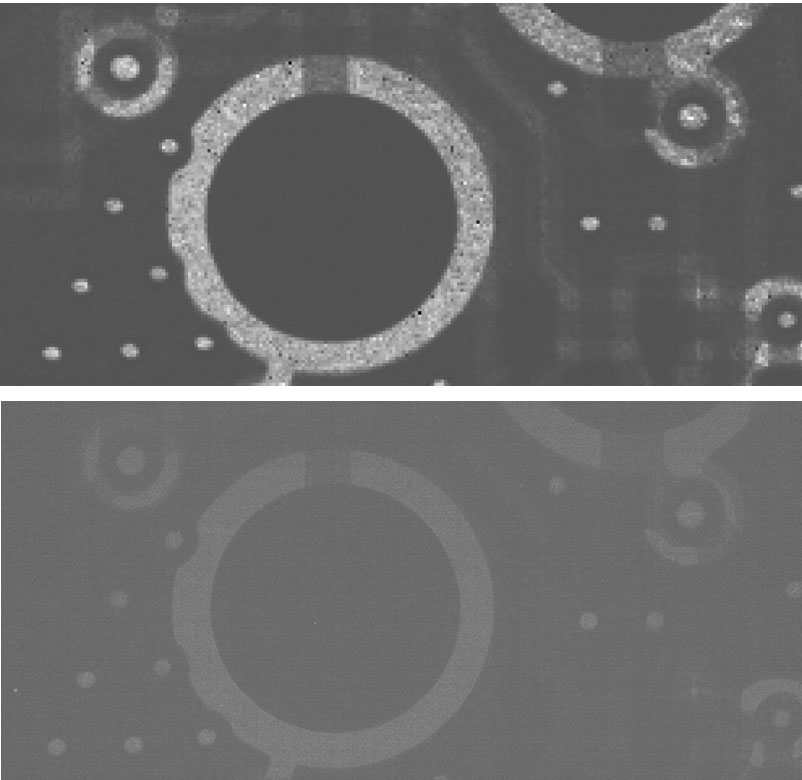
Researchers have demonstrated proof-of-concept clear, high-resolution X-ray images using very little radiation. An image acquired using the new method (a) is compared with conventional radiology (b) under identical photon counts. [Image: Tiqiao Xiao, Shanghai Advanced Research Institute, Chinese Academy of Sciences]
The scientists incorporated ghost imaging—the construction of an image by correlating a spatially modulated reference beam and a signal beam that passes through the image target—into their X-ray approach, which they call “ultra-low-light ghost imaging.” Ghost imaging at X-ray wavelengths has been a hot topic of recent research, but previous research teams have needed to choose between significant drops in the photon count and resolution higher than a few hundred pixels in one direction.
Instead of splitting their source X-ray beam into equal parts, the researchers separated their photons through a Laue diffraction crystal into a weak beam, which went through the sample, and a stronger beam as the reference. Just like astronomers who use a large-aperture “light bucket” telescope to see distant galaxies, the group collected the photons with specialized high-sensitivity detector arrays: one using direct coupling for the sample arm and one with indirect coupling for the reference arm.
Synthetic-aperture reconstruction techniques that use an intelligent algorithm enabled the team to reconstruct a fully resolved image from dozens of measurements, rather than hundreds as in previous X-ray ghost imaging experiments. “Together, these advances balance three key performance indicators simultaneously: a large field of view, ultra-low photon consumption and high imaging resolution,” said Xiao. “This provides a more practical and effective solution for X-ray ghost imaging.”
Testing and next steps
At the X-ray test beamline of the Shanghai Synchrotron Radiation Facility, the scientists compared the new technique to more conventional direct X-ray imaging methods. The new technique produced 1992 × 944-pixel images using less than half of 1% of the photons of existing X-ray platforms. Yet the ultra-low photon technique attained the same contrast-to-noise ratio as traditional imaging.
“Our technology could be combined with routine hospital imaging equipment such as chest X-rays and CT scans,” said Xiao. “It would make medical X-ray imaging safer, which is especially important for children, pregnant patients and people needing frequent scans.”
Next, the researchers will improve the image quality and repeat the experiments with laboratory-based X-ray sources as steps toward clinical medical imaging.
