
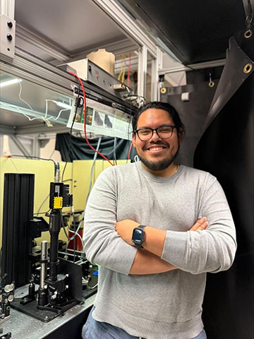
Darwin Quiroz with the laser scanning microscope used for the optical scanning research project. [University of Colorado Boulder]
Researchers at a US university have developed a 2D electrowetting prism scanner that performs well inside a laser scanning microscope (Opt. Express, doi:10.1364/OE.567484 ). The new device could benefit systems ranging from lidar to brain imaging.
Beam steering: from solids to liquids
Multiple types of imaging systems—microscopy, holography, tomography—rely on steering mechanisms to control one or more beams of light passing across a sample. These mechanisms run the gamut from mechanically controlled mirrors and micro-electromechanical systems (MEMS) to acousto-optic deflectors. When miniaturizing such a system for in-vivo usage, such as studies of neurons, scientists must factor in the mass, size and power demands of the steering components.
In electrowetting, an applied voltage modifies the contact angle and the shape of a fluid that conducts electricity. If the fluid is transparent to light, the fluid can act like a flexible lens that can steer the beams passing through it. While electrowetting technology has been used to shape beams in some applications, it’s generally been limited to 1D work.
By actuating the prism near its two resonances, 25 and 72 Hz, the scientists acquired 500 × 500 pixel monochrome images of tiny polystyrene beads.
A 2D electrowetting scanner
A team at the University of Colorado Boulder created an electrowetting prism consisting of four indium tin oxide electrodes surrounding a tube filled with deionized water and a colorless organic liquid with the chemical formula C12H14. The surface tension between the two liquids makes the pairing virtually immune to distortion by gravity, so that only the voltage from the electrodes controls the shape and tilt of the interface between the liquids. A printed circuit board controls the electrodes, which are separated by a 30-μm gap.
“Most laser scanners today use mechanical mirrors to steer beams of light,” study author Darwin Quiroz said. “Our approach replaces that with a transmissive, non-mechanical device that’s smaller, lower-power and potentially easier to scale down into miniature imaging systems.”
After modeling the system on a computer, the group, led by Quiroz, Eduardo Miscles and Mo Zohrabi, experimentally tested the electrowetting prism’s resonance modes with a 633-nm laser. Finally, the team incorporated the prism into a two-photon laser scanning microscope with a 920-nm Ti:sapphire laser as a light source. By actuating the prism near its two resonances, 25 and 72 Hz, the scientists acquired 500 × 500 pixel monochrome images of tiny polystyrene beads.
“Imagine being able to watch brain activity in real time while an animal runs through a maze,” said Quiroz. “That’s the kind of in-vivo imaging this technology could enable, and it could transform how we study neurological conditions like PTSD or Alzheimer’s disease.”
