
Scatterings
Imaging Collagen Fibers
A group of researchers has used intense X-rays to achieve unprecedented resolution of the structure of the protein collagen.
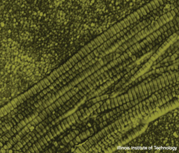
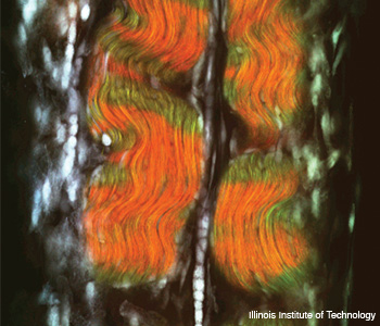 (Top) Collagen fibrils in a collagen fiber, imaged by an atomic force microscope. (Bottom) Collagen fibers (green and red) in a tendon, imaged using second-harmonic generation microscopy.
A group of Illinois Institute of Technology (IIT) researchers has used intense X-rays to achieve unprecedented resolution of the structure of the protein collagen. The research allowed them to see how the enzymes that regulate collagen in connective tissues bind to the molecule (Proc. Natl. Acad. Sci. USA 105, 2824). Their work may provide clues about how collagen becomes disrupted in arthritis and cancer and could eventually lead to drugs that can fight these diseases.
…Log in or become a member to view the full text of this article.
This article may be available for purchase via the search at Optica Publishing Group.
Optica Members get the full text of Optics & Photonics News, plus a variety of other member benefits.
