
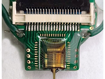
The tip of the probe is about 0.2 mm wide and 0.05 mm thick. [Image: UMass Amherst]
Optogenetics, a method to optically control the activity of neurons using light-sensitive proteins, has become an indispensable tool for neuroscientists. As an alternative to an all-optical approach, optogenetic electrophysiology combines high-resolution electrical recording with optical manipulation of cells. However, up to this point, optoelectronic neural probes could only control brain activity in one direction—excitation or inhibition of neurons—with a single color of light.
Now, researchers at the University of Massachusetts, Amherst, USA, have developed what they say is the first dual-color optoelectronic neural probe (Cell Rep. Phys. Sci., doi: 10.1016/j.xcrp.2023.101702). The device, which allows for bidirectional control of the same set of neurons, could help neuroscientists better understand neurological disorders such as Parkinson’s disease and epilepsy through animal models.
Adding another color
Bidirectionality requires dual-colored light that occupies a broad optical bandwidth, which causes interference when employed alongside optical techniques like fluorescent live-cell imaging. Compared with all-optical methods, optogenetic electrophysiology has the advantage of low crosstalk when using more than one color of light.
Previously, researchers had implemented single-colored microLED-based probes in optoelectronic experiments, which performed well on their own but proved difficult to integrate. So Guangyu Xu and his colleagues aimed to fabricate an implantable neural interface on a single circuit board that could output different colors of light and included arrays of microelectrodes.
"[We wanted to develop] neural probes that can both enhance and inhibit activities of the same neurons via spatially confined dual-color light illumination patterns," said study author Xu, an associate professor of electrical and computer engineering. "In this work, we translated the idea of multicolor LED arrays established in display applications and applied it to the world of neurotechnology."
Better disease models
The researchers chose Chrimson and GtACR2 as the excitatory and inhibitory light-sensitive proteins, respectively, given that their activation spectra are well separated from each other. On the hardware side, they built blue GaN-based microLEDs (462 nm) to activate GtACR2 and red AlGaInP-based micro-LEDs (625 nm) to activate Chrimson.
They implanted the neural probe in the brain of anesthetized mice and were able to excite or inhibit neurons in the mouse somatosensory cortex.
The neural probe contained a total of 16 blue LEDs and 16 red LEDs, with each device being 7 μm × 7 μm in size, along with 17 recording electrodes 20 μm × 20 μm in size. After characterizing its optoelectronic performance, Xu and his colleagues tested their device on mice to assess its capabilities for bidirectional in vivo optogenetic electrophysiology. They implanted the neural probe in the brain of anesthetized mice and were able to excite or inhibit neurons in the mouse somatosensory cortex by illuminating either the red or blue LEDs.
“Applications of these probes include enabling the bidirectional optical dissection of brain circuitry down to cellular levels, which will help functional brain mapping at unprecedented precision,” said Xu. “[They also offer] new tools and assays to establish high-precision neurological disease models, which may ultimately add to the screening of perspective therapeutic interventions.”
