

The SACLA free-electron laser facility in Harima Science Garden City, Japan, where the experiment on the diffraction of ultra-short X-ray pulses on crystalline silicon samples was carried out. [Image: SACLA]
For more than a century, X-ray scattering has been the physicists’ tool of choice for probing the crystalline structure of materials. Modern X-ray free-electron lasers (XFELs) can blast materials with intense femtosecond pulses, but what do the pulses do to the structures they are trying to study?
Using a femtosecond XFEL focused tightly on a silicon nanocrystal sample, an international research team found that X-ray diffraction intensity dropped dramatically when the beam intensity reached a certain threshold (Phys. Rev. Lett., doi: 10.1103/PhysRevLett.131.163201). Simulation of the experimental results revealed just how the first few femtoseconds of the light–matter interaction scrambled the atoms of the material.
Disappearing diffraction signals
Scientists have successfully determined the structure of protein microcrystals with XFEL pulses in the range of 1017 W/cm2. However, many of the studies of the interactions between materials and pulses in this intensity range have been with materials in the gas phase, rather than solids.
Working with the SPring-8 Angstrom Compact free-election Laser (SACLA), part of the SPring-8 synchrotron radiation facility in Japan, the scientists irradiated 10-μm-thick silicon samples with beam intensities at two levels: 2.1 × 10¹⁶ W/cm2 and 4.6 × 10¹⁹ W/cm2. They discovered that when using the higher beam intensities, the resulting diffraction images were up to 50% dimmer than expected.
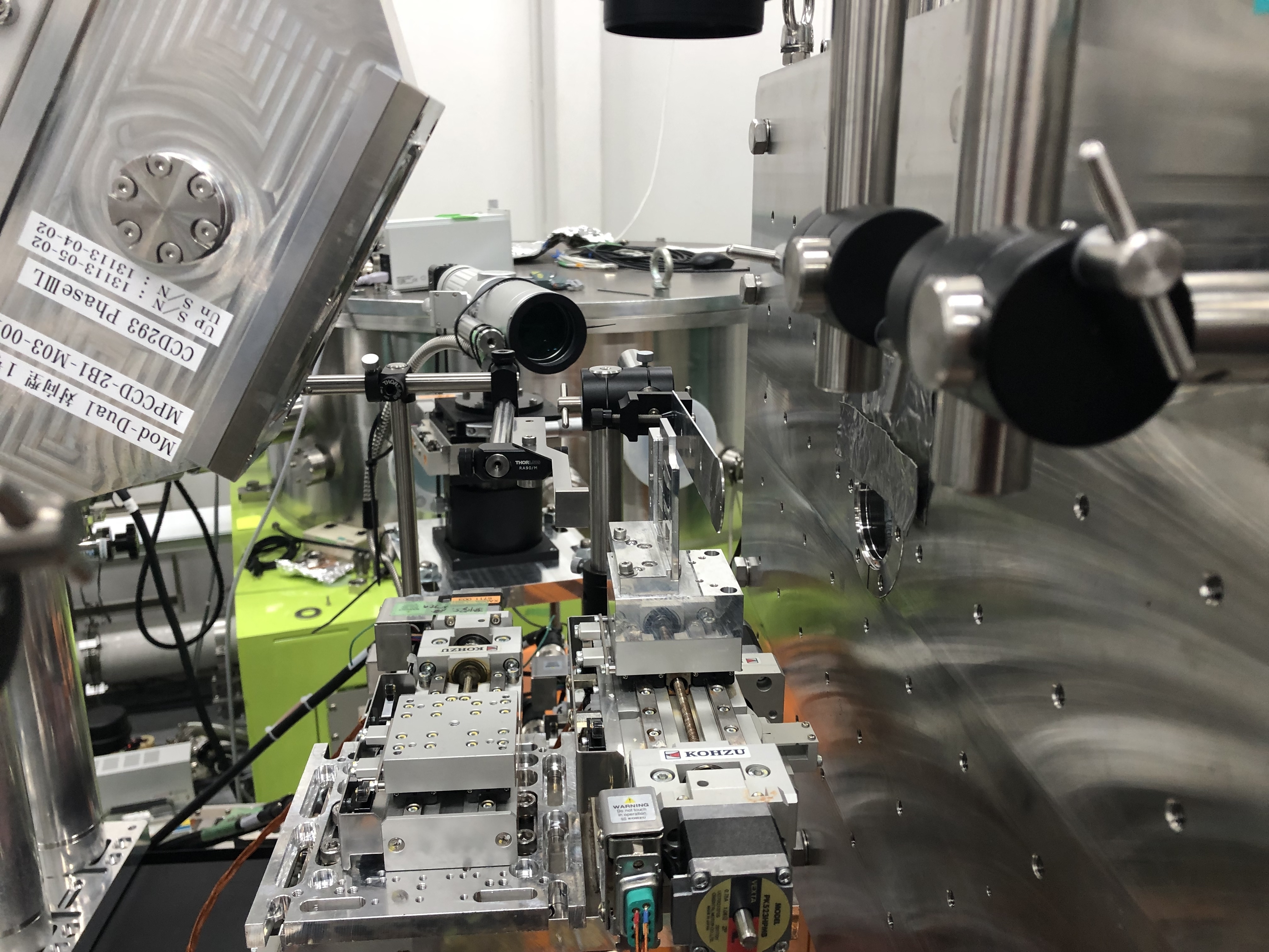
Experimental setup at the SACLA facility for the diffraction experiment on crystalline silicon samples. [Image: SACLA]
“Intuition tells us that the more photons we have, the clearer the diffraction image of the sample should be. This is indeed the case, but only up to a certain X-ray intensity, of the order of tens trillions of watts per square centimeter,” said author Beata Ziaja-Motyka, Institute of Nuclear Physics of the Polish Academy of Sciences, Krakow, Poland and the Deutsches Elektronen-Synchrotron, Hamburg, Germany. “When this value is exceeded … the diffraction signal suddenly starts to weaken.”
Electron knockout
The group, led by Ichiro Inoue of the RIKEN SPring-8 Center, Japan, turned to simulations to explain the paradox. According to the simulations, the blast of high-energy (11.5-keV) photons knocks out not only valence electrons, but also the electrons in shells closer to the atomic nucleus. These deep-shell holes reduce the “atomic scattering factors” that affect the diffraction results. All this ionization occurs within the first 6 fs of the light−matter interaction.
The team hopes that its work will lead to new applications of high-intensity XFEL radiation, such as novel optical devices for pulse-shortening in the X-ray regime. The finding that different atoms respond differently to ultrafast X-ray pulses could also help more accurately reconstruct 3D atomic structures from recorded diffraction images.
