

An artist’s view of an ultrafast laser pulse interacting with the nucleoside molecule uridine. [Image: R. Borrego-Varillas]
A research team from Italy and Germany has used ultrafast laser pulses to shed light on an enduring biochemical mystery: how DNA protects itself from damage by UV radiation from the sun (Nat. Commun., doi: 10.1038/s41467-021-27535-7). The researchers believe that the work, which tracks the biomolecules’ photoprotection mechanisms at picosecond to femtosecond timescales, could prove useful in nanotechnology and pharmacology.
Protecting the genetic blueprint
As the molecules carrying an organism’s genetic blueprint, DNA and RNA need to shield themselves from a continual rain of solar UV photons, the high energy from which otherwise would quickly scramble the genetic instructions. Such scrambling can lead to disorders such as skin cancer. Fortunately, however, these biomolecules feature a remarkable photoprotection mechanism, whereby the excess electronic energy from UV light is dissipated—on timescales as short as hundreds of femtoseconds—to keep the genetic-encoding system free from photodamage.
This ultrafast sleight-of-hand takes place in the nucleosides, molecular units that are among the components from which the backbone of the larger DNA and RNA macromolecules is formed. But while scientists have long known about the robustness of these molecular building blocks, there has been considerable disagreement about the details of how their photoprotection actually works. And—because of the incredibly brief timescales of the process—it’s been a hard problem to crack.
A “real-time movie” of photoprotection
To get at that problem, the researchers behind the new work used ultrafast transient-absorption (TA) spectroscopy to take a “real-time movie”—a series of snapshots of the photoprotection process in action—at a frame rate of less than 30 fs. In TA spectroscopy, a pump pulse resonant with the molecular transition of interest is followed up with a broadband probe pulse, the absorption of which is charted spectroscopically as a function of the pump–probe delay time. This allows the team to track the photoexcitation and subsequent relaxation of the nucleosides and explore the detailed dynamics of the process.
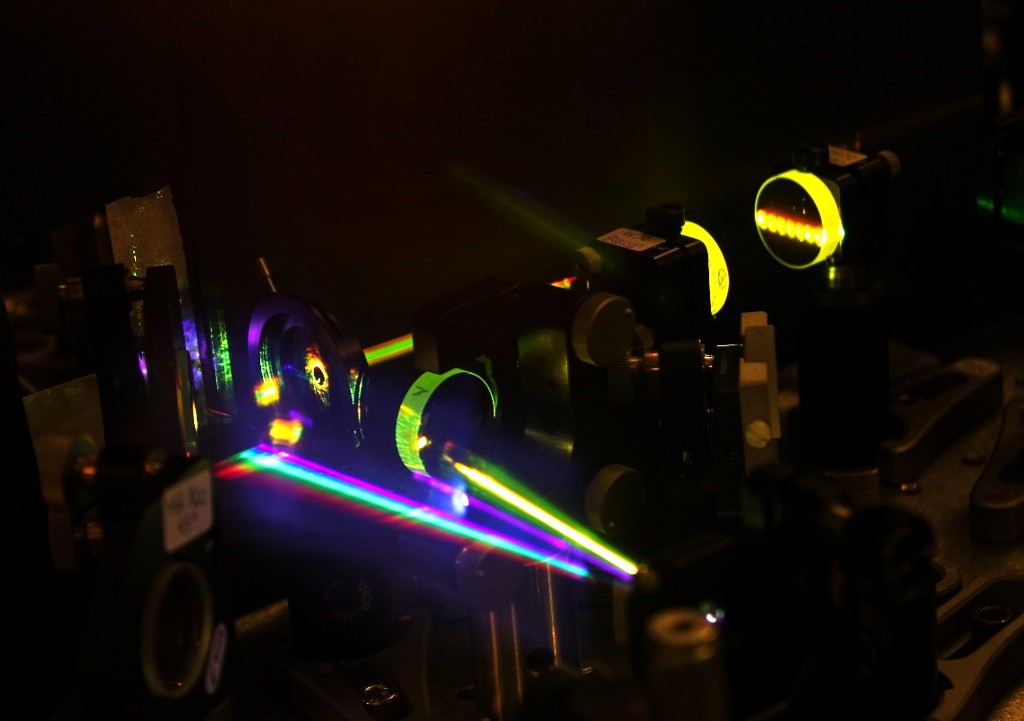
A photo of the experimental setup. [Image: R. Borrego-Varillas] [Enlarge image]
Setting the scene for this particular movie, though, was an laborious process. According to Rocío Borrego-Varillas of IFN-CNR, Italy, a co-first author on the study, it took the team two years to build a beamline that could do TA spectroscopy experiments in the UV range at sub-30-fs time resolution.
The elegant setup resulting from that work begins with a stream of 100-fs pulses from an 800-nm, 1-kH-repetition-rate Ti:sapphire laser, a share of the power from which drives a non-collinear optical parametric amplifier (NOPA). The pulses from the NOPA are then compressed to sub-10-fs duration by a series of chirped mirrors and successively frequency-doubled by nonlinear crystals, to create the UV pump pulses. Broadband probe pulses are created by white-light continuum generation.
Mapping energy dissipation
The team fired pump and probe pulses from this setup on samples of two nucleoside molecules of interest—uridine and 5-methyluridine—dissolved in a phosphate-buffered aqueous solution. The researchers then studied the resulting spectral signatures. By comparing those signatures with numerical simulations of different models, they were able to gain insight on how the nucleosides’ energy dissipation—and, thus, photoprotection mechanism—unfolds on breathtakingly short timescales.
In particular, the ultrafast movie allowed the team to dig into details of nucleoside quantum chemistry that were previously inaccessible. For example, it’s generally been agreed that the ultrafast energy conversion at the heart of the photoprotection mechanism takes place at so-called conical intersections—“molecular funnels” where potential-energy surfaces intersect and rapid photochemistry can take place. But the decay mechanisms by which the photoexcited nucleosides move toward that energetic sweet spot have been unclear.
Spying on the nucleosides
The ultrafast experiments, coupled with the advanced theoretical simulations the team also developed, allowed the researchers to spy on the vibrational signatures of the coherent wave packets as the nucleosides’ energy state moved toward the conical intersection.
For example, the team found that uridine decays to its ground state through a straightforward ballistic process, on timescales of around 100 fs. In contrast, the relaxation of 5-methyluridine lumbers along a multi-step path at a much slower pace of around 1 ps. The team tied that relative sluggishness to the fact that the 5-methyluridine, as its name implies, is dragging along a heavy methyl group not present in the uridine, which increases the inertia and slows down the excited-state decay.
Those details could have practical implications. Study co-first author Artur Nenov, Università degli Studi di Bologna, Italy, observed in a press release accompanying the research that the fact that photoprotection in 5-methyluridine is ten times slower than in uridine “might explain why DNA strands containing thymidine, a nucleoside similar to methyluridine, are more prone to UV photodamage.”
The team hopes that this sort of insight, made possible by ultrafast laser science, could prove worthwhile for applications in nanotechnology and pharmacology. “Understanding how nucleosides interact with light in these very short timescales is a stepping stone to comprehend the complex physical processes leading to DNA photodamage,” Borrego-Varillas said.
