
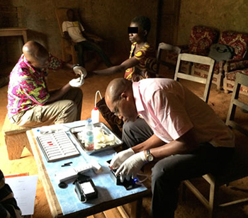
A pilot test of CellScope Loa in Cameroon found that the device was as good as conventional blood smears in detecting the Loa loa parasitic worm. Credit: courtesy of NIAID
Using mobile-phone microscopy for point-of-care diagnostics isn’t new, but incorporating video into the technology is. A team of researchers, led by bioengineer Daniel Fletcher at the University of California, Berkeley, USA, has developed a smartphone video microscope that can quantify parasitic worms from whole blood (Sci. Transl. Med., doi: 10.1126/scitranslmed.aaa3480). The instrument, named CellScope Loa, is the latest mobile microscopy technology from Fletcher’s lab. A pilot study with the Berkeley group and researchers at the U.S. National Institute of Allergy and Infectious Disease (NIAID), France and Cameroon demonstrated the device’s ability to quickly identify Loa loa worm concentration just as accurately as more time-consuming microscope methods.
In Central Africa, infection with parasitic worms Onchocerca volvulus and Wuchereria bancrofti, which can cause blindness and elephantiasis, is highly endemic. Treatment with ivermectin-based drugs is effective, but has been discontinued because it can cause severe and often fatal neurological side effects in people who are co-infected with Loa loa. Therefore, quickly diagnosing a Loa loa infection can greatly reduce a patient’s risk for disease and drug-related death—and could allow resumption of mass administration of ivermectin for those with O. volvulus and W. bancrofti infections.
Currently, health care workers test for Loa loa by drawing a patient’s blood, viewing a prepared and stained sample under a light microscope, and manually counting the moving worms to determine if the parasite load is too high for ivermectin treatment. CellScope Loa promises to provide the same data in about two minutes. It uses a smartphone video camera coupled with a reversed camera lens module for imaging, and a light-emitting diode array for bright-field video microscopy with a resolution of < 6.5 µm over a 4 x 3.16 mm field of view. A custom-algorithm app on the phone quantifies the parasites by measuring the movement of red blood cells (caused by worm “wriggling”) collected in a thin glass capillary.
In the Cameroon pilot study, researchers tested samples from 33 subjects potentially infected with Loa loa. They found that CellScope Loa was 94 percent specific (compared with traditional microscopy) and 100 percent sensitive for patients who had parasite concentrations high enough to cause severe adverse events to ivermectin (30,000 worms per milliliter of blood).
Fletcher says CellScope Loa “demonstrates what technology can do to help fill a void for populations that are suffering from terrible, but treatable diseases.”
