
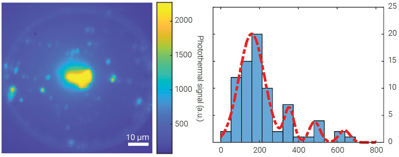
 [Enlarge image]Left: Photothermal map of 5-nm quantum dots on a microtoroid resonator. Right: Intensity analysis revealing single dots and aggregates. [Adapted from Ref. 1; CC-BY-4.0]
Imaging individual particles billionths of a meter across, without fluorescent labels or cryogenic cooling, opens a new window into the smallest structures light can reveal.1 Among these are quantum dots: semiconductor nanocrystals only a few nanometers wide, valued for their roles in quantum information, bioimaging and optoelectronics. Detecting the smallest quantum dots, however, is especially difficult. Surface trap states, tiny imperfections on their surfaces that quench their emission, and their minute size and weak scattering make them nearly invisible to label-free optical methods, which depend on refractive-index changes rather than emitted light.
To overcome these limitations, we combined photothermal microscopy with an ultra-high-Q whispering gallery mode microtoroid resonator, creating an imaging platform with extreme sensitivity and high spatial resolution. Light circulates thousands of times around the microtoroid’s periphery, generating intense optical fields and long photon lifetimes. These properties amplify even minute refractive-index changes to detectable levels, enabling label-free visualization of individual nanoscale objects.
In our setup, a pump laser tuned to the quantum dot’s absorption band is modulated at a fixed frequency. The absorbed light slightly heats the quantum dot, inducing a local-refractive-index change—a photothermal effect. The resulting signal is extracted with a double lock-in detection scheme that integrates the FLOWER platform2,3 with a lock-in amplifier, suppressing noise and achieving a signal-to-noise ratio above 104 for single 5-nm quantum dots. Scanning the toroid point-by-point with the pump beam yields a spatial map pinpointing each dot’s location.
The maps show bright, discrete spots from individual quantum dots, alongside larger features from aggregates. Because the particles are covalently attached to the resonator surface, the images capture a fixed nanoscale landscape. Even scanning electron microscopy struggled to confirm the presence of single particles—small random features, potentially dust, could be mistaken for dots. Our approach allows identification of single emitters and clusters without relying on photoluminescence.
High-resolution photothermal mapping of objects thousands of times smaller than the width of a human hair pushes the boundaries of optical metrology to new frontiers. This approach is poised to impact fields from quantum photonics, where it could enable deterministic placement and quality control of single-photon emitters, to the life sciences, where it could reveal the architecture of viruses and protein complexes in their native environments. Looking ahead, such sensitivity could make it possible to watch the molecular machinery of life operate in real time, capturing the motions of single proteins, viruses and nanoscale devices as they function, advancing how we design materials, diagnose disease and build quantum technologies.
Researchers
Judith Su, University of Arizona, USA
References
1. S. Hao, S. Suebka and J. Su, Light Sci. Appl. 13, 195 (2024).
2. S. Suebka, A. Gin and J. Su, Nat. Protoc. 1, 35 (2025).
3. J. Su, A.F. Goldberg and B.M. Stoltz, Light Sci. Appl. 5, e16001 (2016).
