
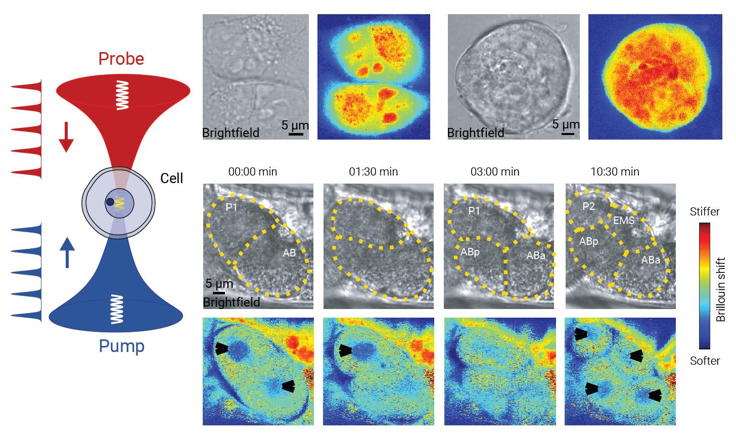
 [Enlarge image]Left: Schematic of the pulsed-laser stimulated Brillouin scattering microscopy. Right: Brightfield and Brillouin images for HeLa cells, a lung cancer tumor organoid and a developing C. elegans embryo from two-cell stage to four-cell stage in vivo.
The mechanical properties of cells and tissues play a critical role in regulating biological activities, including cell fate, tissue morphogenesis and organ function. Brillouin microscopy has emerged as a powerful tool for probing viscoelastic parameters, offering the distinct advantage of noninvasive 3D measurements at diffraction-limited resolution.1–3 The frequency shift and linewidth of Brillouin spectra acquired at the focal spot are directly related to local stiffness and viscosity.
However, the low-scattering cross-section and subtle frequency shifts of biological samples pose significant challenges for achieving rapid spectral acquisition. For measurements requiring both high spatial resolution and spectral precision, existing Brillouin microscopy techniques are limited by long acquisition times—typically on the order of tens of milliseconds per pixel—that restrict their applicability for highly dynamic biological processes.
Recently, we have improved the imaging speed by employing stimulated Brillouin scattering microscopy with a dual-wavelength pulsed fiber laser system.4 The single-frequency 1560-nm seed laser was chopped, amplified and then frequency doubled to generate 780-nm optical pulses with nanosecond pulse width. Synchronized pump and probe pulses are used to excite stimulated Brillouin scattering within the biological sample, precisely at the same focal spot of a pair of opposing objectives.
Our method leverages the low-duty-cycle, high-peak-power laser pulses to enhance the stimulated Brillouin gain efficiency, thereby increasing the signal amplitude. Combined with auto-balanced detection to suppress the intensity noise of the pulsed laser, our approach enhances the signal-to-noise ratio and hence the imaging speed. As a result, we achieved Brillouin imaging of living organisms with a pixel time of 200 μs at an average optical power of 30 mW, while maintaining high frequency precision (7.7 MHz), spectral resolution (132 MHz) and spatial resolution (0.49 × 0.49 × 2.1 µm3). The high temporal resolution of the pulsed-laser stimulated Brillouin scattering microscope allowed us to capture early C. elegans embryonic development in vivo, revealing nuclear dynamics during the transition from the two-cell to the four-cell stage.
We believe that pulsed-laser stimulated Brillouin scattering microscopy has the promise to enable broad applications in mechano-biological research. Future developments in fiber-nonlinearity suppression and noise reduction could further enhance sensitivity and imaging speed, potentially bringing the throughput of Brillouin microscopy on par with that of confocal fluorescence microscopy.
Researchers
Yun Qi, Shuai Yao, Zi-Xuan Du, Jin-Rui Zhang, Cuiyun Zhou, Xiaohu Fu, Wei-Biao Chen and Fan Yang, Shanghai Institute of Optics and Fine Mechanics, China
Huan Li, Ting Mi and Jingjing Xie, ShanghaiTech University, China
Yu-Han Chen, Yu-Fan Wang, Yun Luo, Shi-Qing Cai and Jiu-Lin Du, Insitute of Neuroscience, China
Xuyan He and Peng Xia, Zhejiang University, China
References
1. G. Scarcelli et al. Nat. Methods 12, 1132, (2015).
2. I. Remer et al. Nat. Methods 17, 913, (2020).
3. F. Yang et al. Nat. Methods 20, 1971, (2023).
4. Y. Qi et al. Nat. Photon. 19, 879, (2025).
