
Feature
Optical Tweezers Bring Micromachines to Biology
Optical tweezers, using focused laser beams to manipulate objects on the nano- and microscale, allow exploration of a world of curiosities at the borderline between physics and biology.
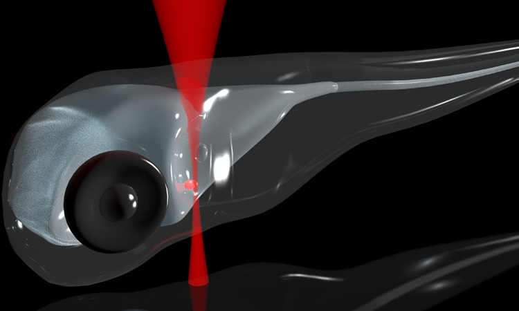 Optical tweezers have been used to trap and manipulate the tiny, 55-μm otoliths, or ear stones, in zebrafish larvae, and to study how vertebrates use those structures to detect and respond to acceleration. [A.V. Kashchuk]
Laser micromanipulation—the trapping, translation and rotation of microscopic particles by focused laser beams—has found worldwide application in basic and applied research. Particularly exciting has been the use of such “optical tweezers” in biology to study phenomena ranging from cell division to DNA bonds to the trapping of blood cells in vivo in the capillaries of mouse ears. Recent work has even extended the technique to micromanipulation of 50-micron-diameter structures in living zebrafish embryos, and to observing the behavioral responses that result.
…Log in or become a member to view the full text of this article.
This article may be available for purchase via the search at Optica Publishing Group.
Optica Members get the full text of Optics & Photonics News, plus a variety of other member benefits.
