
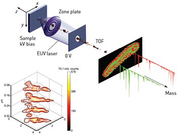
 (Top) Schematic of the EUV MSI method. (Bottom) 3-D composition image showing iso-lines of the distribution of the most intense ion, with 70.1 m/z in the mass spectra, from a single Mycobacterium smegmatis. The brown contours identify the edge of the bacterium. The image is constructed from spline-interpolated 0.3×0.3×0.08 μm3 voxels.
Analytical probes capable of mapping molecular composition in 3-D at the nanoscale will transform materials research, biology and medicine. Mass-spectral imaging (MSI) is one of the most powerful methods to visualize the spatial organization of multiple molecular components on solid samples. However, it is challenging for MSI to map molecular composition in 3-D with submicron resolution.
We have demonstrated a new MSI method that combines extreme ultraviolet (EUV) laser ablation with mass spectrometry to obtain 3-D composition images with nanoscale resolution.1 In the EUV MSI setup, bright laser pulses from a compact 46.9-nm-wavelength laser2 are focused into nanometer-size spots to ablate craters a few nanometers deep on selected regions of the sample. The ions in the laser-created plasma are extracted and identified by their mass-to-charge ratio (m/z) using a time-of-flight (TOF) mass spectrometer. The 3-D composition images are constructed from the analysis of spatially resolved mass spectra obtained as the sample is displaced with respect to the focused laser beam.
The use of λ=46.9 nm laser light for MSI has three unique advantages. First, it can be focused into spots of ∼100 nm, significantly smaller than those obtained with UV light resulting in superior lateral resolution. Second, its absorption depth is extremely shallow— around 20 nm—thus making it possible to ablate craters a few nanometers deep by controlling the laser fluence at the sample. And, third, the EUV photons can directly photoionize organic molecules.
In a recent experiment we showed that EUV MSI could detect singly ionized intact organic analyte ions within the mass range of up to 400 m/z, with a superior sensitivity of 0.01 attomol. We assessed molecular composition across a sharp boundary with a lateral resolution of 75 nm and a depth resolution of 20 nm. We exploited the high localization of the focused EUV light to realize 3-D nanoscale chemical imaging of a single Mycobacterium smegmatis. The composition map in the figure illustrates the content of a salient fragment with 70.1 m/z within the bacterium. Optimization of the interaction of the EUV laser light and organic materials, in combination with post-ablation ionization, will help extend the mass range and maximize the potential of this nanoscale 3-D MSI method for life science applications.
Researchers
I. Kuznetsov, J. Filevich, F. Dong, M. Woolston, W. Chao, E.H. Anderson, E.R. Bernstein,
D.C. Crick, J.J. Rocca and C.S. Menoni, Colorado State University, Fort Collins, Colo., USA
References
1. I. Kuznetsov et al. Nat. Commun. 6, 6944 (2015).
2. S. Heinbuch et al. Opt. Express 13, 4050–4055 (2005).
