

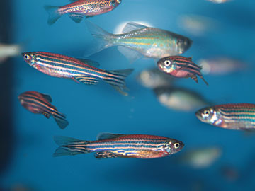
A new imaging system has captured images of a seizure making its way through the brain of a zebrafish larva. [Image: Getty Images / kazakovmaksim]
A seizure is a sudden, intense wave of electrical energy that passes through the brain in a matter of seconds. Now, researchers at the University of Georgia, USA, have devised an imaging system that can track the onset and evolution of these fast-moving events in zebrafish larvae, an animal model that is often used for neuroscience research (Biomed. Opt. Express, doi: 10.1364/BOE.596096).
One slice at a time
The imaging system is based on a technique called light-sheet fluorescence microscopy, which offers a fast and powerful tool for imaging dynamic processes in living creatures. A thin sheet of laser light is used to illuminate a single slice of the specimen at a time, recording the fluorescence emitted from each location to create a high-resolution image without damaging the organism.
Light-sheet microscopy has already been used to capture large-scale neural activity in zebrafish larvae, but almost all previous studies have been restricted to two dimensions. Fast volumetric imaging of other biological processes has been achieved by sweeping the light sheet through the sample and then using an electrically tunable lens to synchronize the focal plane of the imaging objective with the scanned light sheet. But these flexible lenses can introduce significant optical aberrations when used to image large volumes, which has limited their use for studying the global dynamics of the brain.
A synchronized system
Previous work by the University of Georgia team has shown that these aberrations can be corrected using adaptive optics, but the extra data needed to enhance the image quality slowed down the rate of acquisition. To solve that problem, the team has now devised a scheme that synchronizes the settings of the adaptive optics system with the continuous scan of the light sheet. This dynamic process allows the system to capture four full volumes every second—some seven times faster than the original demonstration.
Initial tests with fluorescent beads show that applying adaptive optics in this fast-acquisition mode expands the imaging area that achieves subcellular resolution by a factor of five. When used to image zebrafish larvae, adaptive optics makes the neuronal cells appear more distinct and spherical in shape when compared with the uncorrected images.
The team then used the system to record 3D videos of seizure events in zebrafish larvae. These images reveal that the electrical activity originates toward the back of the brain and then progresses toward the part of the midbrain that processes visual information. The seizure reaches a peak after around 15 seconds, with the electrical energy gradually subsiding as the wave moves toward the front of the brain.
The researchers now plan to use this improved imaging capability to better understand the propagation of electrical waves during a seizure and to probe the neural processes that can cause or inhibit seizure events. They hope that the insights gained from these studies could inform improved treatments of brain diseases and disorders.
