
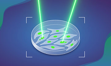
MAGIC spots cells with a particular visible feature, such as the presence of micronuclei, and marks them using a system involving a laser and a photoconvertible dye. [Image: Daniela Velasco, EMBL]
Chromosomal defects can lead to cancer, but detecting faulty cells in tissue samples can be tedious and time-consuming. Researchers in Europe have combined lasers, fluorescent dyes and artificial intelligence to speed up the process of finding cells with abnormal chromosomes and shed light on the formation of cell defects (Nature, doi: 10.1038/s41586-025-09632-5).
The technique, called MAGIC (machine-learning-assisted genomics and imaging convergence), automates the detection of live cells with atypical nuclei in images. An algorithm trained on images of similarly defective cells tags the problematic ones with laser light. Standard cell-separation methods can then isolate the abnormal cells for genomic analysis.
Speeding up the search
In recent years, scientists have learned crucial lessons about chromosomal abnormalities through fluorescence, confocal and phase-contrast microscopy of living cells. However, only a relatively few cells in a given tissue sample may possess these defects at any given time, and they may die off before scientists can manually isolate and study them.
The control script was trained on an annotated dataset to look for cells containing micronuclei—small clumps of chromosomes that broke off and became isolated during previous cell division.
In their effort to speed up the search, researchers at the European Molecular Biology Laboratory (EMBL), Germany, started by preparing two lines of live human cells, one cultured from breast tissue and one from retinal tissue. In each line, the team made the cells susceptible to laser tagging in one of two ways: by introducing a plasmid to induce photolabeling in each cell or by treating the cultures with a tracking dye that fluoresces red under 405-nm light. Neither one of these techniques made the cells more susceptible to chromosomal abnormalities.
Testing the MAGIC system
For these experiments, the EMBL scientists collected images of the cell cultures with a confocal laser scanning microscope running in an automated mode. The control script was trained on an annotated dataset to look for cells containing micronuclei—small clumps of chromosomes that broke off and became isolated during previous cell division. The presence of such structures makes it more likely that a cell will become cancer.
The algorithm then feeds the coordinates of the defective cells to the MAGIC system, which zaps those cells with the blue-light laser to change their color permanently. The photo-induced change allows the system to sort out the targeted cells and isolate them for gene sequencing.
Using MAGIC, the EMBL group found spontaneous chromosomal abnormalities in about 10% of the cell divisions—and the rate nearly doubles when the cells lack p53, a gene known to suppress tumor formation.
