

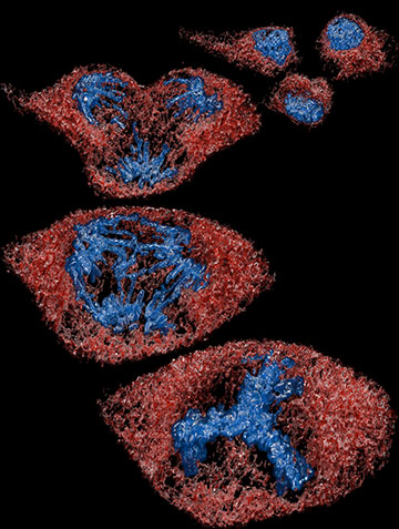
The MOSAIC microscope captures images in five dimensions—the three of space plus time and color—to reveal events that are otherwise hard to view. This image shows a cancerous epithelial cell from a pig dividing into three daughter cells. [Image: Advanced Bioimaging Center/UC Berkeley]
A collaboration based in the United States has combined a dozen different types of optical microscope to create a single device that can image biological specimens from single cells to complete embryos over timescales of fractions of a second to days (Nat. Methods, doi: 10.1038/s41592-026-03066-1). The researchers reckon that the new system, already being put to use by a number of groups around the world, should cut costs in modern microscopy laboratories but caution that they still need to develop machine learning capable of analyzing the terabytes of output data.
Conflicting modalities
Since shortly after its invention in the 17th century, optical microscopy has been used to study cells in a wide range of living systems—including cultured cell lines, flies and larger animals such as mice. The varied studies, in turn, have spurred numerous forms of microscopy, among them widefield and confocal microscopy as well as a range of fluorescence imaging techniques. More recently, techniques have been developed to perform such microscopy at resolutions beyond the diffraction limit.
However, the different modalities often have conflicting requirements that necessitate distinct pieces of hardware, making research more expensive. Lenses with high numerical apertures, for example, boost resolution but limit the field of view, while “light-sheet” microscopes designed to minimize potentially harmful doses of optical radiation can complicate the mounting of specimens. What’s more, the output from virtually all techniques is impaired by optical aberrations generated in the study of multi-cellular organisms.
Five-dimensional imaging
Srigokul Upadhyayula and Eric Betzig at the University of California, Berkeley, along with Wesley Legant at the University of North Carolina at Chapel Hill, have attempted to overcome these problems by building a reconfigurable microscope that they call Multimodal Optical Scope with Adaptive Imaging Correction (MOSAIC). Occupying about 1m3 of bench space, the system comprises numerous carefully laid-out lenses, mirrors, cameras and other optical components. At its core are three objectives that sit alongside the specimen in a temperature-controlled chamber, while its adaptive optics include a deformable mirror shaped by 69 different motors.
By repositioning specific components, MOSAIC can switch fairly quickly between 12 different types of microscopy. These include widefield epifluorescence imaging, three-dimensional structured illumination microscopy, lattice light-sheet microscopy, image scanning microscopy and, for in vivo imaging, two-photon point-scanning microscopy.
By repositioning specific components, MOSAIC can switch fairly quickly between 12 different types of microscopy.
As Betzig puts it, the system is designed to carry out five-dimensional imaging. He explains that to understand what goes on in an organism or perhaps just a single cell it is necessary to study the behavior of a range of structures, down to the sub-cellular or even molecular level. Distinct fluorescent labels allow researchers to track these varied structures—it is the different colors of the labels that constitute the fifth dimension, in addition to the more familiar three dimensions of space and one of time.
The researchers put their system through its paces by carrying out a wide range of measurements on different biological systems. These included tracking single molecules in cultured cells, observing how the cellular sub-units known as organelles restructure, and following neural activity in live mice.
One striking demonstration of the system’s potential, according to Upadhyayula and colleagues, was its ability to image the regrowth of an amputated zebrafish’s tail fin. The researchers recorded a 12-hour video of the phenomenon, revealing, for example, how cells near the wound communicate with one another and fuse together in the healing process and how tiny fibers underneath the skin shift as the process unfolds.
Hurdles to overcome and future directions
The researchers say that since publishing preprints and detailed instructions on how to assemble MOSAIC, more than a dozen other groups have reproduced the system in their own labs over the last six years. They are hopeful that other groups will follow suit given the lower costs and smaller volume of the new device compared with operating single-purpose microscopes separately. They also say that rapid switching between the various modes enables correlations to be made between different images of the same dynamic living system.
However, they acknowledge that a number of hurdles stand in the way of widespread use. For one thing, MOSAIC is complex. Pointing out that its documentation consists of almost 1,000 pages of slides and notes, the researchers say that aligning the numerous modes “requires considerable skill and patience” and that even once aligned operation is only possible following “a substantial investment of time and training”—burdens, they add, that can be lightened if a group decides to commission only some of the modes.
At the same time, the scientists say, there is a need for a new type of artificial intelligence to handle the immense amount of data that MOSAIC produces—up to 4 terabytes per hour or some 30-100 terabytes for each dataset. They note that these data can be processed in real time using a powerful enough distributed computing platform but say that the processed data sets are too big and complex to be analyzed by humans. They argue that one possible solution is to develop a suitable “multimodal machine-learning 5D foundation model,” something the researchers say they are working on but that will not be easy. Betzig described the task as “at the bleeding edge of AI research today.”
Ultimately, assuming they can crack the data analysis problem, Upadhyayula and colleagues envisage MOSAIC being run in centralized facilities akin to modern astronomical observatories. These “cell observatories,” they say, would give biologists access to advanced microscopes and high-performance computers alongside curated data sets and pre-trained models—with expert staff on hand to guide them through the acquisition, processing and analysis of data.
