

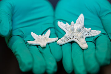
Ling Li holds the skeletons of two differently sized sea stars, both showing the calcium carbonate skeleton. [Image: Bella Ciervo, Penn Engineering]
From the simplest diatoms to iridescent mollusk shells and brilliant bird feathers, the structures built by animals have inspired scientists to develop new and improved materials. Now, researchers based at universities in the United States and Germany have found that an aquatic animal incorporates light-guiding optical structures into its endoskeleton (Proc. Natl. Acad. Sci., doi:10.1073/pnas.2533437123).
The arrays of tiny cone-shaped calcite-type structures found within the skeleton of a tropical sea star collect light over a broad field of view and concentrate it in an internal cavity, suggesting that it has evolved a way to combine mechanical support and optical sensing in the same material system. Although the scientists—who discovered the light guides while seeking inspiration for lightweight yet strong materials—have not yet determined whether the starfish actually see images the way humans experience vision, team members hope that the research could lead to new types of multifunctional materials.
A closer look
The sea star in this study is Protoreaster nodosus, a warm-water echinoderm found in the Indian and Pacific oceans. In life, the starfish appears brownish or reddish with dark protrusions that scare off potential predators. It has garnered the nickname “chocolate-chip starfish” after the cookie or biscuit.
When these creatures die, they leave behind mostly calcium carbonate skeletons that are lightweight and porous, yet strong. Researchers led by Ling Li, a materials science professor at the University of Pennsylvania, USA, and Penn graduate student Liuni Chen noticed lens-like structures on the tips of the star’s five arms. These terminal plates consist of magnesium-rich calcite.
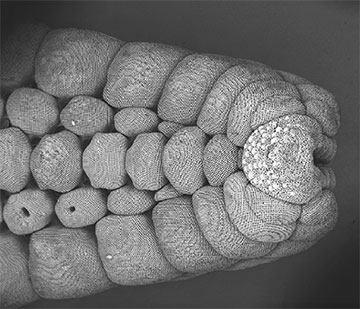
A scanning electron microscopy (SEM) image of the sea star arm skeleton with soft tissue removed. The terminal plate, located at the tip of the arm, contains numerous, bump-like light-guiding structures. The image measures approximately 5 mm in width. [Image: Liuni Chen]
Upon further examination with multiple imaging techniques—microcomputed tomography, energy-dispersive X-ray spectroscopy and diffractive methods from crystallography—the researchers showed that the terminal plates on the arm tips contain conical light-guiding structures up to 100 μm in diameter and 250 μm in length. Multiple such structures on the tips pointed to an internal cavity a couple of hundred micrometers wide.
Shining light on star sight
Shining light through the guiding structures revealed a bright spot beneath the terminal plate, but the researchers found that the array of these structures would nevertheless have several blind spots. This finding suggests that the “eyes” might be suited for sensing environmental brightness changes rather than producing images of external objects.
The light-guiding cones transmit about 70% of the incident light, and each array of cones has a field of view of 120 degrees. Whether the sea stars have actual photoreceptor tissue inside the cavities surrounding the conical “eyes” has yet to be determined.
The research team, which includes scientists affiliated with six other laboratories, hopes that the microstructure design of the sea star skeleton can lead to new, ultralight glass- and ceramic-based materials that are both load-bearing and light-transmitting, for instance, lightweight foams, packaging materials and structural components for spacecraft.
“Imagine a protective panel that not only absorbs impacts but also monitors environmental conditions, detects damage or provides feedback about its surroundings,” says Li. “The sea star demonstrates that such combinations may be possible.”
