
![]()
Marianne Liebi, Meitian Wang and Christian Appel (from left to right) at the Swiss Light Source. [Image: © Paul Scherrer Institute PSI/Mahir Dzambegovic]
A team of researchers based in Switzerland and Sweden has refined a decade-old X-ray diffraction technique to measure the 3D nanoscale information of biological structures (Small Methods, doi: 10.1002/smtd.202500162). Small-angle-X-ray scattering tensor tomography (SAS-TT), originally developed beginning in 2014, combines computer tomography concepts with scanning small-angle X-ray scattering.
To demonstrate the capabilities of SAS-TT, the scientists used the Swiss Light Source (SLS), a synchrotron located at the Paul Scherrer Institute (PSI) in Switzerland, to image the alignment of collagen fibers in one of the small bones of the human ear. “In doing so, we have taken the leap from a scientific method to a practical technique,” said study author Christian Appel, PSI Center for Photon Science, in a press release accompanying the research.
Boosting the scanning speed
Previously, SAS-TT was limited by the time-consuming nature of its measurements, which could take more than 24 hours for a single sample. A typical sample, roughly millimeters in size, required several hundred 2D projections with a few thousand diffraction patterns each.
To this end, Appel and his colleagues used the macromolecular crystallography beamline PX-I at SLS, which features a micro focused X-ray beam with high flux to enable faster and finer scanning of samples. Since it is optimized for high-throughput cryogenic crystallography, PX-I also allowed them to carry out measurements under cryogenic conditions to reduce X-ray-induced heating and radiation damage.
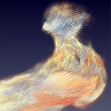
Scientists at PSI were able to observe the local collagen structures in an ossicle by scanning it with an X-ray beam. The different colors of the cylinders indicate how strongly the collagen bundles are spatially aligned in a section measuring 20 × 20 × 20 µm. [Image: © Paul Scherrer Institute PSI/Christian Appel]
“We have now refined the method so that we can record a complete tomogram in just over an hour,” said study author Meitian Wang, beamline scientist at the PSI Center for Photon Science.
Biological applications
The researchers reported a record for a full tomogram measured in 1.2 hours, which is 15 times faster than the first measurements in 2014. They performed a pilot study on a human incus, one of the three auditory ossicles in the middle ear, which measured 2.8 × 1.6 mm2. The imaging revealed the main orientation of the mineralized collagen fibrils and their anisotropy, which could be used to model the human middle ear in more detail.
Recent upgrades to the SLS, revealed in August 2025, will mean even faster measurements. SLS experiments, including those featuring SAS-TT, will benefit from light up to 1,000 times more intense than before.
“Combining a higher resolution with a higher speed of the measurement opens up completely new possibilities for tensor tomography, especially in biomedical applications,” said Appel.
