
![]()
Illustration of the mAxialtrode, showing how it combines light, electrical signals and medication in a single needle-thin tube that can be used to control and measure activity in different layers of the brain and to deliver medication directly. [Image: Kunyang Sui]
When used as neurological implants and probes, soft polymer-based optical fibers produce less inflammation in brain tissue than rigid silicon-based fibers and microdevices. However, most of these softer probes developed over the last decade interact with nerves only at the very tip of the fiber—and don’t reveal interactions between multiple layers of the brain.
Now, researchers in Denmark and the United Kingdom have devised a fiber-based brain electrode with functioning interfaces along the length of the probe (Adv. Sci., doi:10.1002/advs.202519744). Tiny channels within the probe can deliver fluid medications or accommodate thin electrodes for recording neuronal signals. The scientists say the fiber probe, dubbed “microchannel axialtrode” or “mAxialtrobe,” can assist in basic brain research, with clinical applications such as drug delivery or neuronal stimulation further into the future.
Inside the axial electrode
The research team, based at the Technical University of Denmark and including researchers from two UK universities, began with custom-drawn optical fibers containing a 200-μm-wide polycarbonate core and acrylic cladding, specifically designed with eight axial hollow channels inside the cladding just outside the core. The fibers carry light at wavelengths of 470 and 650 nm, chosen because they stimulate many specialized light-activated proteins called opsins.
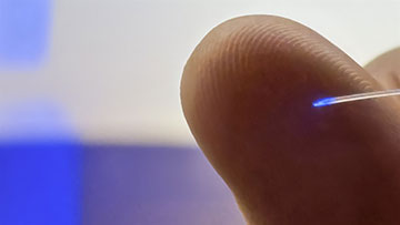
The fiber in the brain implant is less than half a millimeter thick and is so flexible that it moves with the brain instead of cutting through the tissue. [Image: Peter Aagaard Brixen]
For experiments involving electrodes, the scientists placed tungsten wires with a diameter of 20 μm inside the microchannels. The team trimmed the fiber tips at various acute angles rather than cutting them perpendicular to the core, as is done traditionally.
Experimental results
The researchers characterized the light distribution of the angled fiber tips and the fluid delivery capabilities of the microchannels, among other parameters. They then implanted a mAxialtrobe into a living mouse, with some of the probe’s electrodes inside the animal’s hippocampus and others in the cortex. With the application of a suitable bandpass filter, the fiber device detected higher-amplitude theta brain waves in the hippocampus.
Another in vivo experiment demonstrated that the electrodes within the fiber probe could stimulate neural activity within the mouse brain, thanks to the interaction between certain options and blue light.
“Most current brain implants are based on hard materials such as silicon, which can irritate the brain and trigger inflammatory reactions in the tissue,” said Kunyang Sui, Technical University of Denmark. “The new implant differs in that it is made of soft, plastic-like optical fibers and has a specially angled tip that makes it smaller and reduces the damage caused when it is placed in the brain.”
Researchers from the University of Copenhagen, Denmark; University College London, UK; and the University of Manchester, UK, contributed to the study. Future directions for the work include real-time sensing tests and setting the stage for clinical trials of the probe.
