
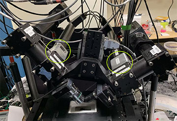
The polarized diSPIM. Liquid crystals used for polarized illumination are shown in green circles. The diSPIM’s dual-view paths meet at a right angle on the sample. [IMAGE: Min Guo]
Measuring the orientation of fluorescent molecules, in addition to their position, can reveal insights about dynamic cell structures. Fluorescence anisotropy imaging has existed for more than half a century, but existing techniques have been constrained to measuring only two dimensions or sparsely labeled single-molecule samples.
Now, researchers in the United States have created a hybrid microscope that can assess the complete 3D orientation and position of ensembles of fluorescent molecules (Proc. Natl. Acad. Sci., doi: 10.1073/pnas.2406679122). The microscope, which combines polarized fluorescence technology with dual-view light sheet microscopy, can record phenomena like 3D protein orientation changes and molecules in the spindle of a dividing cell.
“We see potential applications across cell and organismal biology,” said study author Talon Chandler, CZ Biohub San Francisco, who performed the work as a University of Chicago graduate student. “Our microscope can provide value in cases where the 3D orientation of a fluorescent probe can report difficult-to-access biological information.”
Integrating instruments
Because a large fraction of fluorescent molecules are dipole emitters, polarized light can be used for selective excitation and detection. Biologists then have the opportunity to use optical microscopy to examine a fluorophore’s excitation and emission patterns to draw conclusions about both its position and orientation.
Traditional methods of fluorescence anisotropy imaging are limited to measuring a subset of orientation parameters. More recent single-molecule techniques can assess three-dimensional position, orientation and rotational dynamics—but cannot be applied to ensembles of fluorescent molecules.
Chandler and his colleagues integrated two different technologies—polarized light microscopy and dual-view light-sheet microscopy—to develop a hybrid device that could capture the 3D orientation and position of an ensemble of molecules. In particular, a dual-view light-sheet microscope has two excitation and detection arms that enable diverse illumination and detection polarizations.
“To our knowledge, this microscope is the first to simultaneously measure the 3D position and 3D orientation of fluorescent molecules that densely label a sample,” said Chandler.
The first objective is used to illuminate the sample with a polarized light sheet, while the second objective images the volume.
Validation and the need for speed
The polarized dual-view inverted selective-plane illumination microscope (pol-diSPIM) consists of an asymmetric pair of objectives, each capable of excitation and detection. The first objective is used to illuminate the sample with a polarized light sheet, while the second objective images the volume. Next, the objectives swap roles.
The researchers then repeat these volumetric acquisitions under different polarized illuminations and leverage computational imaging techniques to recover as much information as possible from the raw data. They demonstrated and validated the pol-diSPIM by imaging giant unilamellar vesicles, cellulose and the actin cytoskeleton of cells growing on nanowire grids.
“Our microscope imaged 3D positions and 3D volumes every five seconds, so it is not fast enough to image many of living processes that biologists study,” Chandler said. “Improved variants with faster detection and improved polarization sampling patterns will be valuable.”
