
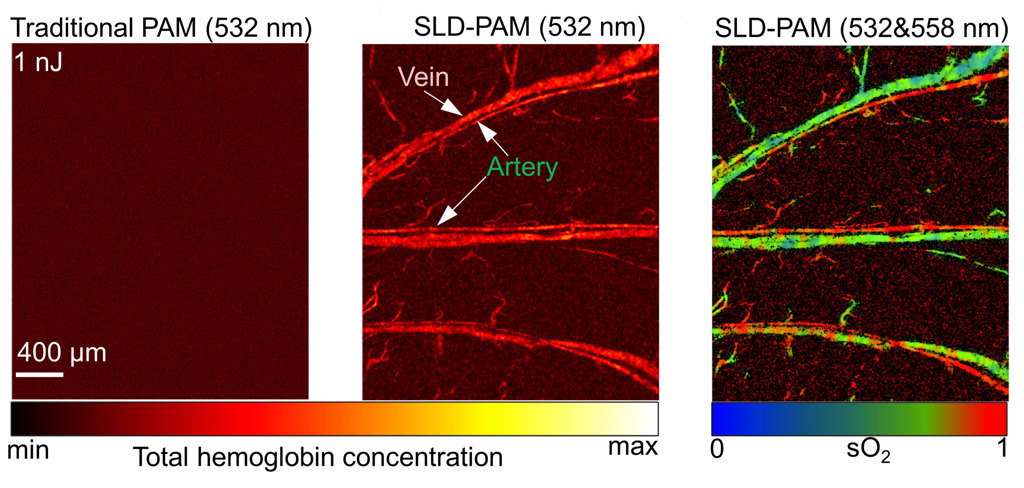
Sound imaging: In these in vivo images of a mouse ear produced at an excitation energy of 1 nJ, the new technique clearly reveals veins and arteries (right) that cannot be seen with traditional photoacoustic microscopy (left) or without the use of the new spectral–spatial filter algorithm (middle). [Image: Y. Zhang et al., doi: 10.1002/advs.202302486] [Enlarge image]
Researchers at the City University of Hong Kong have engineered a photoacoustic microscope that is over 30 times more sensitive than the previous state of the art (Adv. Sci., doi: 10.1002/advs.202302486). The instrument enables high-resolution imaging of live animals at much lower laser powers than has previously been possible, dramatically reducing the risk of damaging biological tissue or its normal function.
Boosting sensitivity with algorithm
Photoacoustic microscopy exploits low-power laser pulses to trigger the emission of ultrasonic waves from light-absorbing molecules within the tissue, which can then be detected and converted into an image. The technique can achieve subcellular resolution at greater depths than conventional optical microscopy, making it a promising method for studying diseases such as cancer, diabetes and stroke, but its limited sensitivity has remained an obstacle to widespread adoption.
To overcome this sensitivity challenge, the researchers combined a series of hardware upgrades with a novel signal-processing algorithm. Their updated photoacoustic probe features a customized acoustic lens with a large numerical aperture, and they also redesigned the optical and acoustic elements to ensure that their focal points are always precisely aligned.
This high-sensitivity probe boosts the collection efficiency of photoacoustic signals by a factor of three, but the biggest improvement was achieved by a new algorithm that suppresses noise and enhances the visibility of weak signals. By combining almost all coherent signals in both the spectral and spatial dimensions, this spectral–spatial filter algorithm maximizes the signal-to-noise ratio and delivers a 33-fold improvement in the overall sensitivity of the system.
Detailed images despite low power
Using a laser excitation energy of 1 nJ—just 1% of the maximum permissible exposure—the technique revealed veins and arteries that cannot be seen without the use of the spectral–spatial filter.
The researchers showed that their updated approach, which they call super-low-dose photoacoustic microscopy (SLD-PAM), can produce high-quality images of microvessels and oxygen saturation in the ears of living mice. Using a laser excitation energy of 1 nJ—just 1% of the maximum permissible exposure—the technique revealed veins and arteries that cannot be seen without the use of the spectral–spatial filter.
Detailed images of the vascular network in both the eyes and brains of living mice were also produced with an excitation energy of just 4 nJ, compared with the tens to hundreds of nanojoules that are typically needed for photoacoustic microscopy. For molecular imaging, tests with a fluorescent dye showed that SLD-PAM can achieve similar image quality with laser pulses that are four times weaker than for a traditional system, preventing photobleaching and increasing the survival rate of the dye from less than 15% to 87%.
“SLD-PAM enables noninvasive imaging of biological tissue with minimal damage to the subjects, offering a powerful and promising tool for anatomical, functional and molecular imaging,” said team leader Lidai Wang. “We believe that SLD-PAM can help advance the applications of photoacoustic imaging, enable numerous new biomedical applications and pave a new avenue for clinical translation.”
