
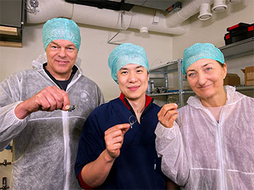
Team lead Edvard Moser, researcher Weijian Zong and team lead May-Britt Moser hold their 2.4 g two-photon brain “miniscope.” [Image: Courtesy of the Kavli Institute for Systems Neuroscience, Norwegian University of Science and Technology]
Scientists use calcium imaging to study brain activity because activated neurons take in calcium ions. Researchers in Norway designed and demonstrated a small two-photon microscope (MINI2P) for large-scale calcium imaging of brain activity in freely moving mice (Cell, doi: 10.1016/j.cell.2022.02.017). The MINI2P weighs only 2.4 grams and features a new electrically tunable lens, super-flexible tapered fiber bundle and an enlarged field-of-view (FOV) that allow for fast, high-quality 3D imaging of different regions of the brain.
The miniscope device and its databases, which are available on an open-access platform, could be used as a collaborative tool to better understand how the brain orchestrates complex activities and how neurological diseases like Alzheimer’s develop.
“Our dream was to invent a window into the brain, so we could see what happens inside when we’re thinking, planning, feeling and remembering,” said group co-lead May-Britt Moser of the Norwegian University of Science and Technology in a press release. “The ability to label different cell types may also enable us to identify which cells are vulnerable to the early changes related to Alzheimer’s disease,” added May-Britt’s research partner and group co-lead Edvard Moser.
Freeing the mice
Researchers use two-photon microscopy to record nerve impulses in the brain by capturing light signals tied to calcium ion intake from fluorescently labeled neurons.
Current two-photon microscopy setups for mouse studies include large benchtop microscopes and tiny head-mounted miniscopes that are attached to an optical imager by a wire tether. Neither design is ideal for recording brain activity in freely moving mice. The benchtop device requires the mouse to remain stationary while reacting to virtual stimuli. The miniscope camera produces low-quality images, and the cumbersome tether restricts the mouse’s behavior.
With the MINI2P, the Moser Lab says it has created a head-mounted two-photon miniscope with the imaging speed and quality of a desktop device and a super flexible tether that does not impede movement.
A tiny cortical observatory
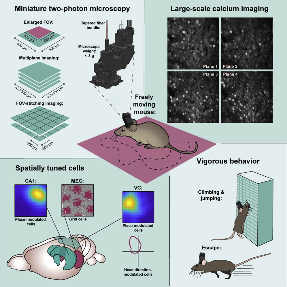
Development of a miniature two-photon miniscope for large-scale calcium imaging in freely moving mice allows stable simultaneous recording of more than a thousand cells across multiple planes of densely active cortical regions in a wide spectrum of behavioral tasks without impeding the animal’s behavior. [Image: W. Zong et al., Cell 185, 1 (2022)] [Enlarge image]
Highlights of the MINI2P design include a new electrically tunable microlens (µTlens) and a super-flexible thin tapered-fiber-bundle (TFB) combined with multi-FOV stitching technology.
The curvature of the µTlens can be quickly manipulated with static voltage, allowing the user to view different focal planes of neuron activity throughout the brain. The µTlens does not generate heat because it operates with minimal electricity, unlike other tunable microlenses that can raise the ambient temperature to a potentially damaging 20°C.
The TFB includes a very thin glass rod that collects and compresses the fluorescence signal from activated neurons. The researchers designed the glass rod to match the signal collection efficiency of traditional fiber bundles while reducing the thickness and increasing the flexibility of the tether.
To expand the MINI2P FOV, the researchers employed multi-FOV stitching—basically, a computer algorithm for quilting together individual high-resolution fluorescence images taken from different areas and depths to get large high-resolution images that can encompass up to 10,000 neurons at once and be used to create 3D maps of brain structures.
Prototype demonstrations and next steps
In proof-of-principle demonstrations, the researchers obtained 2D and 3D images of several mouse brain structures, including the visual cortex, medial entorhinal cortex and hippocampus. They were also able to follow the same brain cells in an active mouse for over a month.
The Moser Lab made the MINI2P open access to allow neuroscience labs anywhere to access blueprints and instructional movies for building their own miniscopes and for sharing their research data. The lab will also provide workshops on using the MINI2P later in 2022.
Edvard and May-Britt Moser were awarded the 2014 Nobel Prize in Physiology or Medicine for discovering grid cells that provide the brain with an internal coordinate system. They co-direct the Kavli Institute for Systems Neuroscience at the Norwegian University of Science and Technology.
