
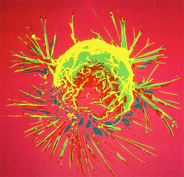
A scanning electron microscope image of a breast cancer cell. [Image: Bruce Wetzel and Harry Schaefer, National Cancer Institute, National Institutes of Health]
Combining near-infrared spectral tomography (NIRST) with contrast-free magnetic resonance imaging (MRI) had been recognized as a non-invasive alternative to some broadly available breast cancer detection and assessment tools. However, reconstructing an image of a tumor after performing MRI-guided NIRST requires considerable time and effort, preventing the technique’s wide dissemination in everyday clinical settings.
To solve the issue, researchers in China, the United States and the United Kingdom have developed a new deep-learning algorithm, Z-Net, that reconstructs the image in a few seconds (Optica, doi: 10.1364/OPTICA.446576). “Z-Net could allow NIRST to become an efficient and effective add-on to non-contrast MRI for breast cancer screening and diagnosis because it allows MRI-guided NIRST images to be recovered in nearly real time,” the research team leader, Keith Paulsen of Dartmouth College, USA, said in a press release accompanying the research.
Merging structural and spectral information
Breast cancer is the most commonly diagnosed cancer in the world. After such a diagnosis, health care providers routinely use dynamic contrast-enhanced (DCE) MRI—one of the most sensitive tools for breast cancer detection—to assess the extent of the disease. But that requires intravenous injection of contrast agent or ionizing radiation, and has a substantial false-positive rate as a screening tool.
In a 2015 clinical study, Paulsen and colleagues found that health care providers could diagnose breast cancer more accurately by performing MRI concurrently with NIRST, which characterizes soft tissue’s optical properties in the 600 to 1000 nm range and provides functional information about the tissue region in question. However, incorporating structural information provided by the MRI into NIRST required segmenting magnetic resonance images into regions with and without tumor or modeling the light propagation of tissue—both time consuming and prone to errors.
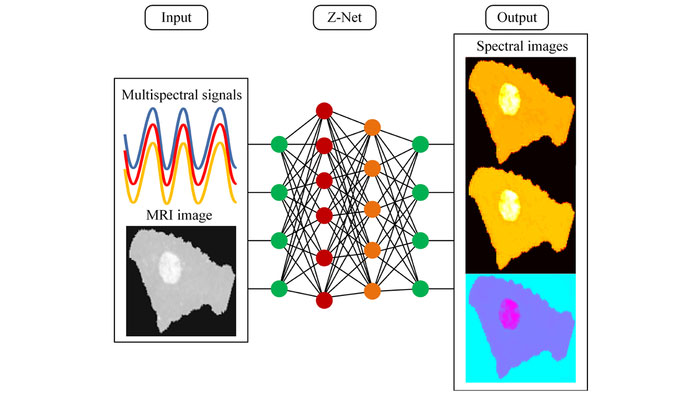
Z-Net deep-learning algorithm can combine spectral and MRI data to reconstruct images within seconds. [Image: Keith Paulsen, Dartmouth College] [Enlarge image]
The researchers developed their new deep-learning algorithm, Z-Net, to get rid of those needs. Deep learning is a form of machine learning implemented in artificial neural networks that are thought to mimic some aspects of the brain. Once trained, these networks can shine in a variety of complex tasks such as image classification and reconstruction.
As input, Z-Net takes MRI images and optical signals at nine different wavelengths between 661 and 948 nm. The algorithm then spits out reconstructed images for chromophore concentrations of three species: oxy-hemoglobin (HbO), deoxy-hemoglobin (Hb) and water.
Reconstruction in seconds
After the researchers trained Z-Net using datasets of simulated data—created using 16 light-source and detector pairs uniformly distributed around the circumferences of 82-mm-diameter circles—the algorithm could reconstruct images in a few seconds. Using simulated data instead of real-patient data for training also helped the researchers save time, said Paulsen.
The researchers fed another set of simulated data into the algorithm after training to confirm that the qualities of reconstructed images were up to par. They then applied Z-Net to MRI-guided NIRST data collected from two women with undiagnosed abnormalities. The reconstructed images from the two breast exams provided enough information to distinguish between a malignant abnormality and a benign one, a finding later confirmed by biopsies.
Even though deep learning had been used in optical image reconstruction before, the researchers write that this is the first time deep learning was applied in combined multimodality image reconstruction.
In addition to potentially enabling NIRST to become a routine add-on to MRI for breast cancer screening and diagnosis, Z-Net “can also be readily adapted for use with other cancers and diseases for which multimodality imaging data are available,” said Paulsen. The researchers are working to expand Z-Net to 3D patient data and larger clinical studies.
