
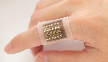
A team at the University of California, San Diego (UCSD), has developed a photoacoustic sensor for mapping hemoglobin. The team believes the soft, wearable device could help clinicians diagnose tumors, organ malfunctions and more. [Image: X. Gao for the Jacobs School of Engineering, UCSD]
An interdisciplinary team based at the University of California, San Diego (UCSD), USA, has created a soft, wearable skin patch capable of measuring hemoglobin in deep biological tissues (Nat. Commun., doi: 10.1038/s41467-022-35455-3).
The device, roughly the size of a postage stamp, employs photoacoustic imaging to map hemoglobin under the skin’s surface in 3D. The technology may offer clinicians and researchers another tool for continuous, real-time, noninvasive monitoring of biomolecules with high spatial resolution.
Deep-tissue sensing
While working in the field of soft wearable electronics, Sheng Xu noticed that existing systems had a significant drawback. They could sense signals only in superficial tissues — either on the skin’s surface or directly below the skin. As a result, Xu aimed to build devices with skin-like mechanical properties specifically for deep-tissue sensing.
“There are a lot of more interesting and critical activities in deep tissues and central organs,” said Xu, a UCSD associate professor of nanoengineering. “Those deep signals are believed to have a stronger and faster correlation to disease prognosis and diagnosis.”
Xu and his colleagues designed and fabricated a soft wearable patch based on photoacoustic imaging. Essentially, the device integrates an array of 240 ultrasonic transducers and 24 high-power vertical-cavity surface-emitting laser (VCSEL) diodes at 850 nm in an elastomeric polymer substrate. The patch is only 2.0×1.6 cm and 1.2 mm thick.
The VCSEL diodes emit light pulses that penetrate deep into biological tissue, where hemoglobin molecules absorb part of the energy and convert it into heat. The transient thermoelectric expansion generates acoustic waves, which are received by the piezoelectric transducers. The transducer signal is beamed to software that reconstructs the spatial mapping of the wave emitters—that is, the hemoglobin molecules.
Toward a safe, continuous bedside monitor
The researchers first performed optical, thermal and acoustic characterizations of the soft photoacoustic patch, followed by in vivo validation experiments on the hand, foot, thigh and forearm. For the latter tests, the device successfully captured 3D images of hemoglobin that clearly displayed the vein structures of different body locations before, during and after cuff occlusion. The penetration depth in the in vivo tests was measured at more than 1.1 cm.
The penetration depth in the in vivo tests was measured at more than 1.1 cm.
Xu and his colleagues think the technology might eventually find use as a long-term bedside monitor for patients with life-threatening conditions such as malignant tumors, organ dysfunction, cerebral or gut hemorrhages, and other conditions.
“Other methods, like magnetic resonance imaging or X-ray, are not suitable for continuous monitoring,” said study author Xiaoxiang Gao, a UCSD postdoc. “Ultrasound is a good candidate for point-of-care and is easy to access, but it cannot differentiate between hemoglobin and other components.” Gao added that the patch developed by the team “has a low form factor, is suitable for long-term monitoring, and is safe.”
