
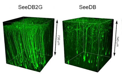
Confocal imaging of a mouse cerebral cortex using SeeDB and SeeDB2 optical clearing agents. Credit: RIKEN
A research team from Japan has demonstrated a new method for obtaining super-resolution 3-D images of neuron networks in brain tissue samples up to a depth of more than 100 µm—10 times deeper than standard imaging methods. The process involves using an agent to make the brain tissue “transparent” by matching the refractive index (RI) of the prepared sample with that of the immersion oil used in fluorescent microscopy (Cell Rep., doi: 10.1016/j.celrep.2016.02.057).
The researchers, led by RIKEN Center for Developmental Biology’s Takeshi Imai, quantitatively tested this new method on two types of brain samples and were able to resolve neuron synapses—areas where two nerve cells meet and communicate with each other—and neuron dendrites. Neuronal mapping, the researchers say, could help researchers better understand normal and abnormal brain function.
Fluorescence microscopy is commonly used in brain imaging because fluorophores can be used to label specific proteins or genes found only in certain brain structures or cells. Optical clearing agents can make brain tissue samples transparent for low-resolution imaging of large structures in the brain. However, current formulations are not ideal for high-resolution 3-D imaging of sub-cellular structures because they can damage brain tissue and disrupt the subcellular structure, or create spherical aberrations which lead to inaccurate images.
To bring these sub-cellular structures into focus, Imai and his colleagues turned to a fructose-based clearing agent that had worked well for them in the past, called SeeDB (shorthand for “See Deep Brain”). They reformulated the agent, appropriately renamed SeeDB2, to clarify fixed brain samples without damaging the tissue. During quantitative tests on mouse and fly brain tissue samples, they found that SeeDB2 was compatible with the bright fluorophores needed for sub-cellular imaging, and also minimized spherical aberrations and maintained fine tissue structure.
