
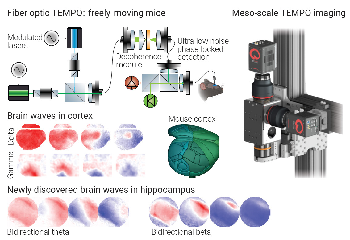
 [Enlarge image]Two TEMPO optical platforms revealed cortical delta and gamma traveling waves and discovered bidirectional theta and beta waves in the hippocampus.
Brain waves are rhythmic changes in neuronal membrane voltage linked to perception, memory and movement.1 For decades, scientists mainly detected brain waves using electrical recordings such as electroencephalography (EEG), which average signals from many neuron types. Optics is transforming this field. Fluorescent genetically encoded voltage indicators (GEVIs) translate millivolt changes in neuronal membrane potentials into shifts in photon flux, enabling voltage recordings from specific cell classes in living brains.2 Two major hurdles have remained, namely, capturing the full frequency range of cognitively relevant rhythms, and doing so across large brain regions, especially during natural, freely moving behavior.
Now, our new optical strategies called TEMPO (transmembrane electrical measurements performed optically) have overcome these barriers, offering shot-noise-limited sensitivity, rejection of blood-flow artifacts, and neuron-type specificity for both imaging and fiber-based recordings.3
A key methodological advance is the use of referenced measurements, whereby a spectrally separable reference signal is recorded alongside the fluorescent GEVI signal at the same tissue site and computationally unmixed. This removes two blood oxygenation–driven nuisances—fast, heartbeat-linked fluctuations and slower shifts from neurovascular coupling—which, while underpinning neuroimaging techniques such as fMRI, are artifactual here. Dual-color acquisition of fluorescence signals using synchronized, low-noise hardware yields a clean, temporally precise readout of membrane voltage.
One technological innovation is a leap in sensitivity for fiber photometry. Lasers provide stable, low-noise, modulated excitation, and lock-in detection reduces electronic noise. However, a tethered animal bends the multimode fiber implanted into the brain, creating interference-induced intensity fluctuations that correlate with behavior. Custom photonics solutions now allow fiber photometry to span DC-120 Hz of neural bandwidth, preserving fast oscillations even as mice move and learn. Another advance is a system that images nearly the entire dorsal neocortex in mice with high spatial and temporal resolution. Custom optics, sCMOS cameras and low-noise LED excitation, combined with computational processing, deliver near-shot-noise-limited performance over unprecedented scales and bandwidths.
These systems provide a unique view of traveling brain waves in the cortex. When applied to the hippocampus, they uncovered previously unknown phenomena: bidirectional theta (3–7 Hz) and beta waves (15–30 Hz), and cross-frequency coupling within single neuron classes.
Demonstrated advances in neuroscience ultimately stem from careful optical design, and the application of photonics in brain research holds great potential for further discoveries. Here, large-scale voltage imaging can reveal how brain waves arise, transmit information and go awry in disease. Optical data about the dynamics of specific neuron types can inform our understanding of EEG, guide diagnostics and therapeutic strategies for neurodegenerative diseases, and inspire AI models.
Researchers
Radosław Chrapkiewicz and Simon Haziza, Stanford University, USA
Mark J. Schnitzer, Stanford University and Howard Hughes Medical Institute, USA
References
1. G. Buzsáki and A. Draguhn, Science 304, 1926 (2004).
2. M.Z. Lin and M.J. Schnitzer, Nat. Neurosci. 19, 1142 (2016).
3. S. Haziza, R. Chrapkiewicz et al. Cell 188, 4401 (2025).
