
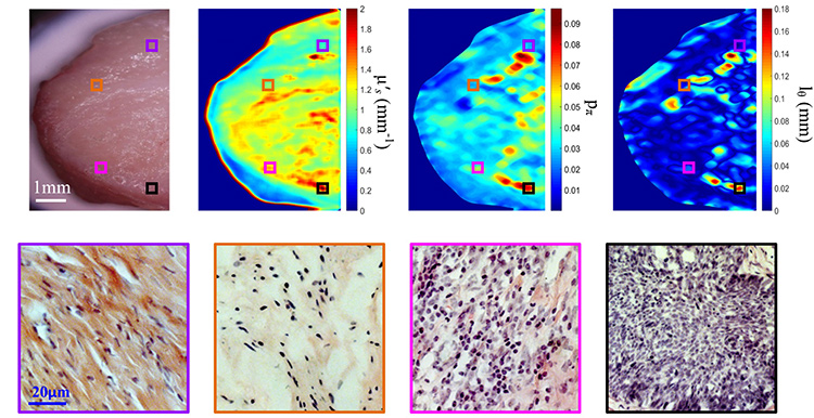
 Top, left to right: Photograph of cervical carcinoma tissue specimen; retrieved μ′s map; retrieved pπ map; and retrieved lΘ map. Bottom: H&E stained histological images for the four highlighted regions in the top row: normal cervical tissue (violet), slight inflammation (orange), severe inflammation (pink), and invasive squamous-cell carcinoma (black). [Enlarge figure]
A central goal in light’s application in biology and medicine is noninvasive diagnosis of the structure and function of tissue. Minimally invasive endoscopic optical imaging, such as confocal techniques, has emerged as an invaluable diagnostic tool for tissue examination, with sensitivity to the subwavelength features. But wide-field imaging requires cumbersome raster scanning that is often beyond the budgets of clinics—and, in any event, wide-field imaging across a field of view of 1 cm2 or more usually can’t resolve tissue microstructure.
The morphological details and microscopic structure of a random medium are encoded in the medium’s phase function, which describes the angular distribution of scattered light upon a single interaction with the medium. When light is backscattered from turbid media, the sensitivity to the phase function and sub-wavelength structures can be increased by using sub-diffusive light at a close source-detector separation, or through a high spatial modulation. This has attracted great interest,1 but remains hampered by the lack of a suitable model relating the reflectance of sub-diffusive light to the explicit form of the phase function and by the degradation of the signal-to-noise ratio for the modulated reflectance at high frequencies.
We have addressed these two limitations2,3 and have developed high spatial frequency domain imaging (HSFDI)4—a noncontact imaging modality that spatially resolves the phase function and maps the tissue’s microscopic scattering structures over a large field of view (greater than 1 cm2). In the accompanying figure, displaying an example of imaging a fresh cervical tissue specimen, it is remarkable that the phase function parameters (pπ, lΘ), unlike the reduced scattering coefficient μ′s, can distinguish cancer, severely inflammatory states, and other disease states with high reliability (p < 0.01).
The steady increase of the backscattering probability pπ and the spreading length lΘ from normal cervical tissue, slight inflammation, severe inflammation, to invasive squamous-cell carcinoma quantifies the enhancement in the background refractive-index fluctuation (“pudding”) and the shrinkage of the size of the prominent scattering centers (“plum”).4,5
Our study demonstrates the ability of HSFDI to rapidly assess whole-tissue specimens for microstructural alterations, in cases where point-source or microscope-based tissue interrogation methods would have to randomly sample a large number of locations to achieve similar robustness. HSFDI may thus fulfill the unmet need for wide-field microscopic structural biomarkers and find clinical applications in rapid screening and diagnosis of tissue.
Researchers
Weihao Lin and Bixin Zeng, Wenzhou Medical University, Wenzhou 325035, China
Min Xu, Hunter College of the City University of New York, N.Y., USA
References
1. D.M. McClatchy et al. Optica 3, 613 (2016).
2. M. Xu. Sci. Rep. 6, 22535 (2016).
3. M. Xu et al. AIP Adv. 6, 125208 (2016).
4. W. Lin et al. Biomed. Opt. Express 9, 2905 (2018).
5. M. Xu. Biomed. Opt. Express 8, 2879 (2017).
