
Feature
Expanding OCT: Quanitifying the Cornea’s Optical Performance
Optical coherence tomography, a well-known method for imaging the retina, may allow physicians to perform quantitative metrology of the eye’s own optics.
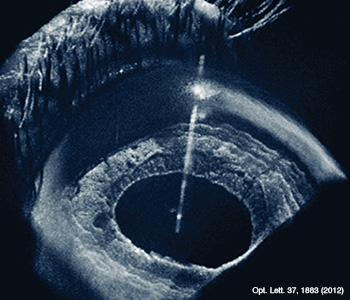 Swept-source OCT allows for simultaneous imaging of the cornea, iris and retina in living patients.
Laser refractive surgeries such as laser in situ keratomileusis (LASIK) and photorefractive keratectomy (PRK) have brought better vision to tens of millions worldwide. But these techniques also exact a cost: because they work by reshaping the eye’s cornea, they make accurate measurement of the cornea’s optical parameters after surgery unreliable using conventional tools. That’s important, not only because inaccurate measurements can lead to suboptimal outcomes in cataract surgery and other procedures, but because, more broadly, improved biometry of the cornea—the eye’s upstream gateway—is a prerequisite for accurate measurement of all of the eye’s other optical elements.
…Log in or become a member to view the full text of this article.
This article may be available for purchase via the search at Optica Publishing Group.
Optica Members get the full text of Optics & Photonics News, plus a variety of other member benefits.
