
Feature
Optical Microangiography: High-Resolution 3-D Imaging of Blood Flow
Using Fourier domain optical coherence tomography, researchers have developed a new method for producing 3-D images of microvascular blood flow.
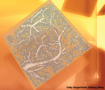
There are many instances when doctors need to visually monitor their patients’ microvascular blood flow—including when they are diagnosing or treating cancer, heart disease and other conditions. Over the past decade, a number of technologies have emerged to meet this need, but each one has limitations. Some traumatize the tissue in a way that may seriously disturb the circulation, for example; others have practical limitations in clinical use. The ideal method of monitoring microcirculation should be noninvasive, versatile and easy to use.
…Log in or become a member to view the full text of this article.
This article may be available for purchase via the search at Optica Publishing Group.
Optica Members get the full text of Optics & Photonics News, plus a variety of other member benefits.
