
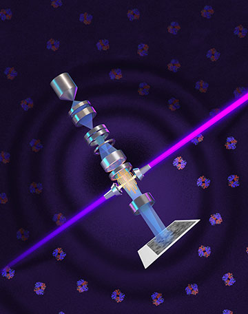
By amplifying a laser beam (purple) and focusing it to a sharp point on an electron beam (blue), researchers have shifted the phase of the latter to increase its phase contrast and thereby image smaller proteins than has previously been possible. [Image: Sayo Studio]
Scientists in the United States have used an extremely intense continuous-wave laser to carry out phase-contrast imaging with an electron microscope (Science, doi: 10.1126/science.aeh0665). They were able to increase the microscope’s resolution when reconstructing the structure of tiny proteins, and they say that the new scheme opens the door to imaging a range of biomolecules too small to study with current techniques.
Enhancing contrast
For almost a century, biologists have used phase contrast to boost the resolution of conventional optical microscopes. While proteins and cellular structures scatter light to generate image contrast via the light’s intensity, that signal is usually too weak to be useful. Phase contrast exploits the fact that the scattered light is slowed down in cells and its phase is shifted. Converting the phase modulation into amplitude modulation involves shifting the phase of the nonscattered light by 90° and focusing the two beams so that they interfere on the observer’s retina.
It has long been researchers’ aim to enhance the contrast of electron microscopy in a similar way. But using physical phase plates to manipulate beams of electrons has proved problematic—often either slashing a beam’s intensity, rendering the resulting images unstable or actually lowering the microscope’s resolution.
The particular technique studied in the latest work is cryo-electron microscopy (cryo-EM), which involves freezing samples in order to limit the damage caused by electron-beam heating. The method has significantly improved scientists’ ability to study the structure of cells and biomolecules, but its full potential has still to be reached. It could theoretically reveal the structure of particles as small as 17 kilodaltons (kDa; 1 dalton being 1/12th the mass of a carbon-12 atom), but to date even proteins weighing in at 50 kDa are tricky to reconstruct.
Preserving power
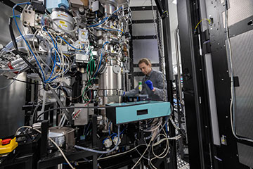
Biohub engineer making adjustments to the laser phase plate cavity inside a cryo-electron microscope. [Image: Dale Ramos/ Biohub]
In 2010, Holger Müller and Robert Glaeser at the University of California, Berkeley, proposed shifting the phase of electron beams without sapping their power by exploiting a “laser phase plate.” The idea is to focus an exceptionally intense continuous laser beam onto the electron beam in order to induce a 90° phase shift in the latter via stimulated Compton scattering. However, doing so requires overcoming a series of technical challenges that many other researchers at the time considered too formidable.
The laser phase plate involves building up the intensity of a continuous-wave laser by reflecting the beam back and forth between the concave mirrors of a Fabry-Pérot cavity thousands of times. This requires making the mirrors from materials lossless enough to prevent appreciable heating despite the laser’s great intensity while polishing the mirrors so that they have “atomic-level smoothness.” The mirrors must also be aligned to within a thousandth of a degree, while the laser and electron beams have to be lined up so that they converge on a spot just a few tens of nanometers wide.
Müller previously built such a system using a fairly old electron microscope, but that device presented a number of limitations—including a wide spread of beam energies and significant electron beam aberration—that in turn capped resolution. He and colleagues at Berkeley have now improved on those results using a more modern instrument that corrects for spherical aberration. They combined this with a 12-W, 1064-nm laser beam that they multiplied to 80 kW in the cavity and focused the beam down to a spot size of 6.7 µm, yielding a record-breaking continuous light intensity of around 400 gigawatts/cm2.
Improved resolution
The researchers demonstrated the power of the new setup by using it to image six samples of small proteins—three containing the protein aldolase from rabbit muscle and three with human hemoglobin (which carries oxygen in blood). Comparing the results against those obtained with the laser switched off, they found that the laser phase plate improved resolution in all six cases. The improvement, they report, was greater for hemoglobin, which has a mass of 64 kDa, than it was for aldolase, which instead weighs 157 kDa.
Impressive as the results are, Müller and colleagues believe there is plenty of room for improvement. Working with engineers at Biohub in Redwood City, CA, they have also developed a laser phase plate with dual lasers, each of which operates at about half the power of the single-beam device in order to lower aberrations. According to Müller, this dual-laser setup brings the microscope’s performance even closer to the theoretical ideal and, he says, should facilitate cryo-electron tomography, which involves imaging proteins not in isolation but within cells, potentially shedding light on biomolecular interactions that cause disease.
