
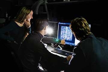
ISTA researchers (left to right) Julia Lyudchik, Mojtaba Tavakoli and Johannes Danzl review a detailed image of the hippocampus produced using LICONN. [Image: ISTA]
An international team of researchers has devised an experimental protocol that exploits a conventional optical microscope to reveal the intricate architecture of the brain (Nature, doi: 10.1038/s41586-025-08985-1). As well as resolving the structure of individual nerve cells, the new experimental method can trace the synaptic pathways that transmit information between neurons and visualize the molecules that play a key role in brain function.
Mapping the brain
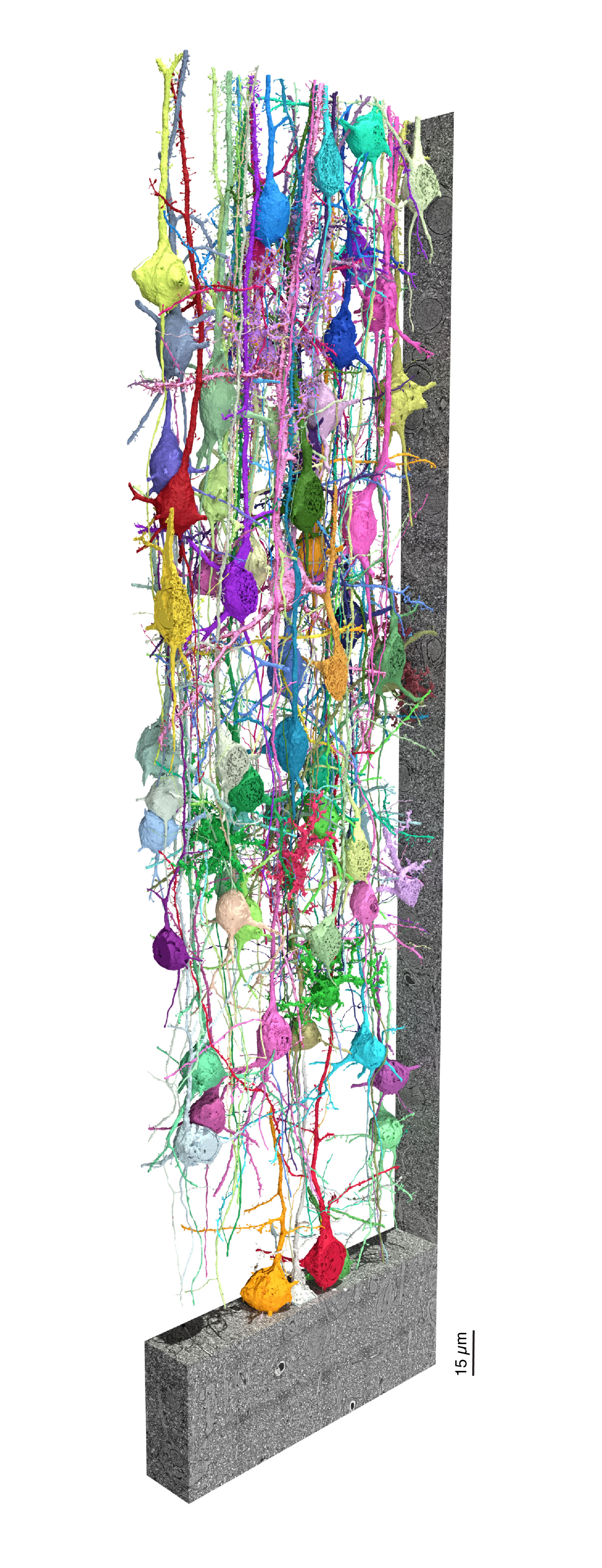
Intricate architecture of a neuronal network. The image shows a 3D rendering of example cells from the mouse primary somatosensory cortex—a brain region that processes touch and body position sensations—produced with LICONN. [Image: Tavakoli, Lyudchik et al./Nature]
While light-based microscopy offers the ideal tool for imaging and identifying specific molecules within the brain, it lacks the resolution to image the fine structure of densely packed brain tissue. The cellular networks that connect neurons can only be reconstructed with electron microscopes, but they are unable to combine these detailed structural mappings with the molecular information needed to study the signaling between cells.
To enable optical microscopy to map out the connectivity of the brain, the researchers developed an experimental pipeline, called LICONN, that they demonstrated using tissue samples taken from mice. They first enhance the imaging resolution by embedding a small piece of the brain tissue inside a hydrogel. The cellular components within the tissue are anchored to the polymer network of the hydrogel, and then water is added to expand the gel. As it swells, the gel gently pushes the cellular structures apart while preserving their spatial arrangement. The researchers developed an iterative process to enable a second stage of expansion, which is key to their method’s success. “This method enhances the effective resolution by a factor of 16, achieving a resolution better than 20 nm,” explains Mojtaba Tavakoli of the Institute of Science and Technology Austria (ISTA), Austria.
A better view
The team showed that the resulting images could reveal individual neurons and subcellular structures, and they could also be used to manually trace the connections between neurons. Once the reliability of the mappings had been established using this manual process, deep-learning techniques were used to automatically identify the neurons and surrounding structures in larger volumes of brain tissue. Combined with the high resolution of the imaging data, this automated process achieves the same accuracy as state-of-the-art techniques based on electron microscopy.
The team then used the microscope to visualize and identify specific molecules, such as synaptic proteins that are involved in sending signals between neurons. Adding this molecular information to the structural data made it possible to place specific molecules within the tissue’s architecture, with deep learning again providing a viable strategy for mapping the location of molecules over larger volumes.
By following this experimental pipeline, the team was able to create 3D images of brain tissue that integrate the structural and molecular information into a single, comprehensive view. “LICONN brings us a step closer to assembling the puzzle pieces of the mammalian brain and better understanding its functioning both in health and disease,” concludes ISTA researcher Johannes Danzl.
