
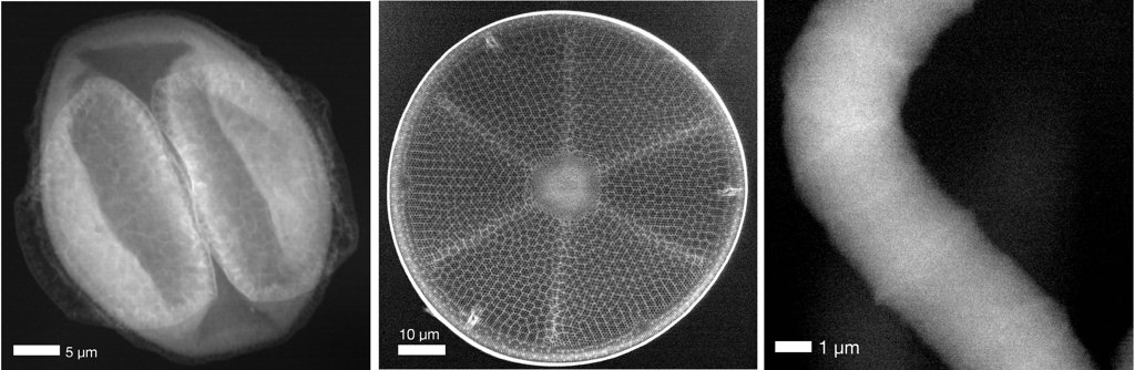
From left to right: a pollen grain, a diatom shell and a cyanobacterium, all imaged using Compton X-ray scattering microscopy at PETRA III. The micrographs could be taken without causing damage to the original samples by using high-energy photons that were highly focused by novel custom lenses. [Image: DESY/Center for Free-Electron Laser Science] [Enlarge image]
X-ray microscopy can produce high-resolution images of tiny objects, but high doses of energetic X-rays can also degrade delicate biological materials. A microscopy technique using a novel multilayered lens has reportedly enabled scientists at a synchrotron radiation laboratory to capture images showing fine details of dried organisms without damaging them (Light Sci. Appl., doi: 10.1038/s41377-023-10076-5).
The technique, devised by researchers in Germany, also relies on a new method for measuring X-ray wavefronts and large-solid-angle pixel-array detectors. It heralds a novel way to examine biological specimens with high-energy beamlines at less than 1% of their X-ray damage threshold.
Balancing wavelength and damage
Choosing the energy level for imaging biological matter is a delicate balancing act involving damage thresholds, desired resolution and other optical considerations such as depth of field. Blasting a sample with 200-keV electrons works only if it is thinner than 0.6 μm. Soft X-rays are primarily absorbed by the atoms in the target materials, prompting electrons to spring out of the atoms and cause damage. Phase-contrast imaging techniques with photons in the 10-keV range penetrate more than soft X-rays and can focus better than the highest-energy beams.
For the latest experiments, the team led by Optica Fellow Saša Bajt and Henry N. Chapman, both of the Deutsches Elektronen-Synchrotron (DESY) used a 60-keV beamline with 0.02-nm wavelength at PETRA III, DESY’s powerful storage-ring synchrotron in Hamburg, Germany. The high energy level permits the use of Compton scattering, which deposits much less energy into the atoms of the material.
To accomplish this task, Bajt and her colleagues devised wedged Laue lenses consisting of alternating layers of silicon carbide and tungsten carbide.
To be useful, though, 60-keV rays need focusing—and they don’t refract well. To accomplish this task, Bajt and her colleagues devised wedged Laue lenses consisting of alternating layers of silicon carbide and tungsten carbide; one such lens is only 35 μm in the direction of beam propagation. Bajt says the Laue lenses are “the culmination of almost a decade of developments, including creating a lab-based X-ray optics testing station.”
“For Compton microscopy, we needed to prepare lenses for very high photon energies, but we could not test them at their operating energy until we set up the microscope at the synchrotron,” she adds. “Another key challenge for Compton microscopy was ensuring all background signals were as low as possible, which required a lot of shielding and precise instrumentation.”
A bright future
The DESY team tested their method on several samples—a grain of pine pollen, a diatom shell and a spirulina bacterium—and achieved a resolution of 70 nm for each. When the researchers compared their pollen images with those of a similar sample obtained with a conventional coherent-scattering imaging method at an energy of 17 keV, their new technique achieved a similar resolution with 2000 times lower X-ray dose.
When the researchers compared their pollen images with those of a similar sample obtained with a conventional coherent-scattering imaging method at an energy of 17 keV, their new technique achieved a similar resolution with 2000 times lower X-ray dose.
The samples in the latest experiments were simply dried and fixed to a substrate. Bajt and her collaborators will experiment with other fixation methods to see what works best for future samples. “Fast cryogenic cooling of the sample may provide the best protection from radiation damage when we approach the highest resolution of 10 nm or so,” she notes.
When DESY brings its next-generation synchrotron, PETRA IV, online—probably in the late 2020s—Compton X-ray microscopy will get another boost. “In scanning microscopy, the name of the game is source brightness,” Bajt says. “With a bright source, we can focus more photons into a smaller spot, allowing us to achieve higher resolution and faster imaging. At our high photon energy of 60 keV, PETRA IV will have an increase in brightness of 1000 times as much as we had here.”
