
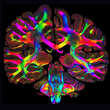
A new imaging technique can map out the orientation and organization of tissue fibers at the microscale. The colors represent different fiber orientations. [Image: Marios Georgiadis/Stanford]
Scientists from the United States, Germany and the Netherlands have demonstrated a computational imaging technique that exploits scattered light to visualize the intricate networks of fibers within human tissue (Nat. Commun., doi: 10.1038/s41467-025-64896-9). Originally designed to study the neural pathways that transmit signals through the brain, the fast and low-cost imaging method can also map out tissue fibers in other parts of the body.
A new and improved method
Microscopic fibers within the body’s connective tissue play a vital role in maintaining the strength and function of bones, organs and muscles, and almost all diseases have a damaging effect on these critical fiber pathways. Existing techniques for imaging tissue structures require specialized equipment and specific protocols for sample preparation, and they can also fail to deliver detailed structural information for fibers that are tightly interwoven.
In contrast, the new technique, called computational scattered light imaging (ComSLI), can produce high-resolution images of specimens created using any preparation method, including those that have been preserved and stored for decades. The only equipment needed is a rotating LED lamp and a standard microscope camera, which record the light scattered from the sample at different angles. Most of the light is scattered perpendicular to the main axis of the fibers, allowing the light patterns to be translated into a color-coded image that maps out the orientation and density of the fibers with micrometer resolution.
Testing efficacy and diverse applications
In tests with brain tissue prepared using various techniques, the method revealed the densely interconnected fiber networks that are typical of a healthy patient.
In tests with brain tissue prepared using various techniques, the method revealed the densely interconnected fiber networks that are typical of a healthy patient. For comparison, a sample taken from a patient with Alzheimer’s disease showed a clear deterioration in the integrity and density of the fiber pathways, with one of the main routes for carrying memory-related signals into the brain becoming barely visible.
The team even used the ComSLI method to image a specimen of brain tissue that had been prepared as long ago as 1904. The technique was still able to reveal the fiber connections within the tissue, offering a new way for researchers to study historical specimens and to trace the lineage of hereditary diseases.
To demonstrate the versatility of the technique, the researchers also produced images of tissue samples taken from muscles, bones and the vascular network. These images revealed distinct fiber patterns related to their physiological roles, such as collagen fibers in bone that follow the lines of mechanical stress and layered fiber structures in the tongue that support movement and flexibility.
“This is a tool that any lab can use,” said Michael Zeineh of Stanford University, USA. “What excites me most is that this approach opens the door for anyone, from small research labs to pathology labs, to uncover new insights from slides they already have.”
