
Researchers used photoacoustic microscopy to image stents through skin. They were able to visualize stents with fractures and compression and other clinical scenarios. [Image: Christoph Burgstedt/ Science photo library/ Getty Images]
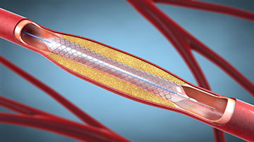
Stents are small mesh tubes made of metallic alloy or polymer used to prop open an artery partially blocked by plaque. Placing a stent during an angioplasty procedure can increase blood flow to the heart and help reduce the chance of a heart attack.
Now, researchers based in China have reportedly discovered that photoacoustic microscopy can noninvasively image a stent through the skin as a way to monitor stent placement for any issues (Opt. Lett., doi: 10.1364/OL.564778). The team says the approach offers advantages over current techniques, which are invasive or use ionizing radiation or contrast agents.
A noninvasive alternative
Both short-term and long-term monitoring of stent status is critical for identifying complications such as stent compression, fractures and malapposition. However, traditional techniques such as X-ray angiography, X-ray fluoroscopy, X-ray computed tomography (CT) and intravascular ultrasound have drawbacks. X-ray-based imaging methods use ionizing radiation, while intravascular ultrasound requires local anesthesia and an incision to place a catheter into the artery.
“We thought that photoacoustic imaging could be used for stent monitoring without the disadvantages of conventional technologies,” said study author Myeongsu Seong, Xi’an Jiaotong–Liverpool University. “Photoacoustic imaging works by picking up acoustic waves that are created when certain materials absorb light and release the energy from the light.”
Seong and his colleagues say they have demonstrated for the first time the capability of photoacoustic imaging—specifically, acoustic-resolution photoacoustic microscopy (AR-PAM)—for monitoring a stent in different conditions through the skin. The work was performed in ex vivo conditions with mouse skin, both label-free and noninvasively.
Seong and his colleagues say they have demonstrated for the first time the capability of photoacoustic imaging ... for monitoring a stent in different conditions through the skin.
Stent scenarios and future directions
The AR-PAM system consisted of an optical parametric oscillator-based wavelength-tunable pulsed laser (670 nm and 1210 nm), beamsplitter, photodiode and a 50-MHz flat ultrasound transducer with an acoustic lens. Tested scenarios included fractures, compression, movement of overlapped stents and deposition of plaque—mimicked by butter—after stenting.
“One of the most interesting results is that we could easily differentiate between the butter we used to mimic a lipid plaque and the stent,” said Seong. “Because plaque and stents absorb light differently, using two wavelengths helped us distinguish them.”
The researchers hope to develop photoacoustic imaging as a complementary tool for frequent stent monitoring to minimize any potential problems that may occur after a placement procedure. Next, the team aims to perform more validations using AR-PAM—particularly in in vivo conditions—to optimize the tradeoff between imaging depth and resolution for different locations.
“Our work was performed in ex vivo conditions,” said researcher Sung-Liang Chen, University of Michigan–Shanghai Jiao Tong University Joint Institute. “Before bringing photoacoustic imaging into clinical noninvasive stent monitoring, in vivo animal experiments and preliminary clinical experiments should be performed, and optimization of system parameters should be done for different application sites.”
