
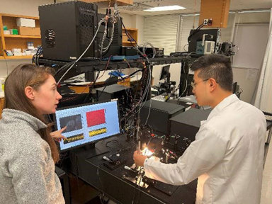
Team members Deirdre Scully and Tian Xia next to the experimental setup for a new OCT-based approach that can directly image the coordination of motile cilia in their natural environment. [Image: Michaela McCown, Baylor College of Medicine]
Motile cilia—tiny, hairlike structures that cover some epithelial cells—contribute to the proper operation of many systems in the human body. Cilliopathies, or abnormal formation or function of cilia, can cause serious health complications, but cilia’s small size has made imaging their dynamics a challenge. The traditional method uses high-speed video microscopy on exposed ciliated surfaces, which requires dissecting the organ—a process that disrupts the cilia’s environment and can lead to inaccuracies in physiological studies.
Now, researchers from Baylor College of Medicine, USA, and Stevens Institute of Technology, USA, have reportedly developed a novel OCT method that enables visualization of the coordination of motile cilia in vivo (Optica; doi: 10.1364/OPTICA.499927). Their approach relies on spatiotemporal mapping, within OCT images, of the phase of intensity fluctuations caused by the cilia’s rhythmic movements.
OCT captures cilia rhythm
One of cilia’s essential jobs in humans and other mammals is to move fluids and particles along the epithelial walls of organs like the fallopian tubes, lungs and brain. They accomplish this task through periodic, coordinated beating that produces so-called metachronal waves propagating along the epithelial surface that transport the substances.
OCT has been gaining popularity as a tool for cilia study, thanks to its ability to conduct high-speed, depth-resolved microscopic imaging with a depth of 1–2 mm in a scattering medium, such as a tissue. Cilia are below the resolution of traditional OCT methods, but their beats create speckles in OCT images that can reveal information about the frequency and phase of the beating at corresponding pixels.
Mapping speckle
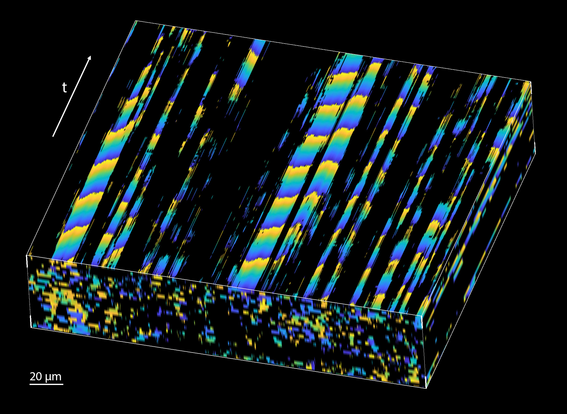
The propagation of the cilia metachronal wave in space and time as captured with the new method. [Image: Tian Xia and Irina V. Larina, Baylor College of Medicine]
The researchers’ new image processing method finds pixels with significant periodic components and then spatiotemporally maps the phase of the dominant frequency, which corresponds to the cilia beat frequency. This both allows direct visualization of the metachronal-wave propagation and provides its velocity.
The team validated the approach using bright-field video microscopy, and then used it to explore the effect of temperature on metachronal-wave velocity in extracted, intact mouse fallopian tubes. Since the researchers were able to image through tissue layers without disrupting the fallopian tubes’ environment, they were able to confirm for the first time that the velocity of the metachronal wave increases with temperature.
They also successfully performed in vivo imaging of cilia metachronal waves within the mouse fallopian tube to reveal wave propagation in any direction. They write that this ability to image cilia dynamics in a living organism is a major advantage of the OCT-based metachronal wave analysis.
Research and clinical applications
The researchers say that, in addition to providing new insights into the female reproductive system, their technique could be modified to study cilia in a variety of organs. They also note that the method has the potential to be used for diagnostic purposes if it is integrated with fiber-based OCT probes that can be guided through the fallopian tube, which are currently in clinical trials.
“Our new method gives access to this previously inaccessible physiological process,” says research team leader Irina V. Larina, Baylor College of Medicine. “We are excited to see what it will reveal about specific cilia roles in female reproductive physiology and specific links between ciliopathy and infertility.”