
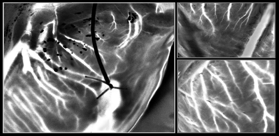
Laser Speckle Orthogonal Contrast Imaging captures details of blood vessels in a beating heart outside the body. [Image: Plyer et al., doi: 10.1117/1.JBO.28.4.046007] [Enlarge image]
For a heart transplantation to be successful, screening donated hearts for congenital abnormalities is essential. A preservation technique called ex situ heart perfusion (ESHP) allows doctors to observe the performance of these hearts outside the body and exclude the ones with issues. However, conducting invasive coronary angiography—the so-called gold-standard method for diagnosing coronary artery diseases—during ESHP can damage the heart and lead to a transplant failure.
Now researchers in France report using an optical technique to image coronary blood circulation in donated hearts during ESHP (J. Biomed. Opt., doi: 10.1117/1.JBO.28.4.0I cr46007). By applying a speckle imaging technique and a postprocessing method, the researchers say they could observe vascular microstructures down to the order of 100 µm in real time in a beating heart.
Imaging and processing
Coronary angiography is a procedure that uses contrast agents and X-rays to visualize the blood flow in the arteries. While the method is favored for its high contrast and spatial resolution, it can also cause heart complications during ESHP due to the accumulation of contrast agents, tears in artery walls and infections.
For an alternative approach, the researchers tapped a previously developed method called Laser Speckle Orthogonal Contrast Imaging (LSOCI), which uses a polarimetric filtering process that prevents surface scattering from reaching a camera and therefore ensures that speckle patterns that vary with time are mainly generated by the multiple scattering of moving red blood cells. In the latest paper, the researchers applied this technique to observe the small blood vessels of a cardiac muscle.
The team first mounted a 785-nm laser and a polarimetric camera on an articulated arm so that the devices would hover over a pig’s heart that was being perfused with oxygenated blood. The researchers kept the heart beating at 80 beats per minute using an external pacemaker. They then used the laser and camera to generate and image rapidly varying speckle patterns.
To be able to observe blood vessels while the heart was beating, the researchers further improved LSOCI with a method called Multi-Period-Enhanced Signal-to-Noise Ratio (MPE-SNR). They first recorded a series of images of the beating heart for several cardiac cycles. Then for the frames with the same and neighboring temporal positions within each cycle, the researchers weeded out frames where the heart and its vessels had moved too much. As a result, by only considering the frames that survived the process, the researchers could create a new sequence that visualized the vasculatures with a high signal-to-noise ratio.
Wide dissemination
At the end, the combination of LSOCI and MPE-SNR allowed the researchers to noninvasively image the complete peripheral vascular system of a beating heart with high spatial resolution within a few seconds.
The researchers write that the simplicity of the experimental setup should help its wide dissemination for a variety of surgical procedures and for monitoring other phenomena in the human artery system that exhibit periodic blood flow.