
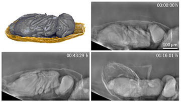
Researchers in Germany have developed a new technique that can capture detailed images of living organisms with low X-ray doses, which they used to track tiny parasitoid wasps for over half an hour as they emerged from their host eggs. [Image: Rebecca Spiecker, Karlsruhe Institute of Technology]
X-ray imaging provides detailed views of the insides of organisms and other objects that are opaque to visible light. However, damage to biological tissue limits the doses of X-rays that can be applied to living creatures―a particular problem in the case of high-resolution imaging.
Now, scientists in Germany have reportedly shown how even the tiniest insects can be observed over long periods of time, by using X-ray optics and single-photon detectors to improve the dose efficiency of phase-contrast imaging (Optica, doi: 10.1364/OPTICA.500978). They achieved almost 100% efficiency at micrometer resolution, up to two orders of magnitude better than more conventional scintillator-based detection.
Avoiding damaging doses
Phase-contrast imaging involves measuring differences in phase accumulated by electromagnetic waves traveling through regions with very slight mismatches in refractive index. This is particularly useful when imaging soft tissue, which provides little variation in the absorption of incoming light.
Researchers have already shown how phase-contrast imaging can be used to study living organisms at resolutions of better than a micrometer. But the need to avoid damaging doses―flux density having to be particularly high at such tiny scales―has restricted such imaging to short timeframes. Typically, samples can be probed for no more than a few minutes.
Imaging can be done on longer timescales by maximizing the fraction of an X-ray dose used to create the image. However, boosting this dose efficiency is complicated by the need to magnify the image when operating at high resolutions.
In conventional setups, incoming X-ray photons are converted to longer-wavelength visible light by a scintillator. These visible photons are then guided by an optical microscope to a pixel-array camera. However, a given (high) resolution requires the microscope to have a certain depth of field, which limits the scintillator's thickness. This in turn puts a cap on the probability of intercepting any given photon, limiting the detector's efficiency.
An additional headache comes from the process of forming an image by converting phase shifts into measurable intensity contrasts. The microscope has an incoherent optical transfer function that impedes image formation, in particular at high frequencies―which are those needed for high resolutions.
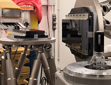
The new X-ray imaging technique uses a much lower X-ray dose thanks to two Bragg magnifier crystals (center) and a single-photon-counting detector (on the left). The sample is shown on the right. [Image: Rebecca Spiecker, Karlsruhe Institute of Technology]
X-ray magnification
In the latest work, Rebecca Spiecker and Tilo Baumbach at the Karlsruhe Institute of Technology, alongside colleagues at a number of other institutes in Germany, have shown how to overcome this problem by using optics to magnify X-rays directly. They did so with what is known as a Bragg magnifier, which consists of two crystals of silicon, each cut so that its outer surface is oriented at a specific finite angle with respect to the crystallographic planes. This leads to asymmetric diffraction and significant magnification of the X-ray image.
The crystals together magnify a monochromatic X-ray beam in both the horizontal and vertical directions. This direct magnification of the original beam allows the X-rays to be detected by very efficient large pixels in the form of a single-photon-counting detector made from a 500-µm-thick high-Z material (gallium arsenide).
The researchers put their system to the test by connecting it to the PETRA III synchrotron storage ring at the Deutsches Elektronen-Synchrotron (DESY) laboratory in Hamburg and comparing its performance with that of a detector system featuring a 12-μm-thick scintillator made from lutetium oxyorthosilicate. Using X-rays with 31KeV of energy, they reached detection efficiencies of about 91%, which, combined with an excellent optical transfer function, yielded quantum efficiencies for the high-resolution image components more than two orders of magnitude higher than those of the conventional setup.
Zooming in on tiny insects
What's more, Spiecker and colleagues showed that they could film a very small insect in vivo over extended periods. The animal in question was a parasitic wasp known as Trichogramma cacoeciae, which infests the eggs of a wide range of crop pests and as such is a valuable agricultural tool. The researchers found that they could expose wasp-containing eggs to lower-dose X-rays for over half an hour without any ill effects. The study yielded footage of a wasp emerging from its egg over the course of more than an hour and showed that these insects can move their mandibles independently of one another and so penetrate a variety of eggshells.
The researchers argue that in addition to in vivo imaging of small animals, their technique could also enable prolonged in situ studies in materials science―probing, for example, the response of polymers to mechanical loads. First, however, they aim to improve their setup by using bigger crystals and larger single-photon counters as well as enhancing mechanical stability. They also plan to make their technique available to users at PETRA III.
