
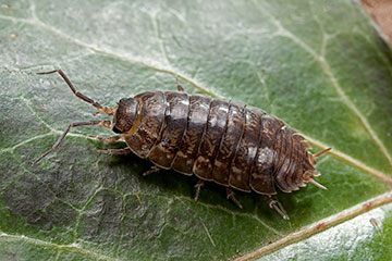
Woodlice sequester metallic particles inside their digestive organ, potentially transferring the contamination to other animals in the local ecosystem. [Image: P. Starosta / Getty Images]
Scientists at Cardiff University, UK, have devised a new imaging method for assessing the toxicity of metallic nanoparticles in biological organisms (Appl. Phys. Lett., doi: 10.1063/5.0140651). Such tiny particles often leach from industrial manufacturing plants into the surrounding environment, but researchers currently lack the tools needed to understand their effects on local habitats and wildlife.
The study centers on the common woodlouse, which is able to ingest and process organic matter containing metal contaminants. These small and ubiquitous creatures have a specialized digestive organ, called a hepatopancreas, that allows them to store and eventually expel metallic particles—but also offers a route for metal deposits to be transferred to other animals in the food chain.
Detecting gold nanoparticles
The optical technique devised by the Cardiff team detects the location and distribution of individual gold nanoparticles inside the hepatopancreas. “By using gold nanoparticles, which would not normally be present in the woodlice diet, we can study the journey of nanoparticles inside complex biological systems,” commented team member Wolfgang Langbein.
For conventional optical methods, however, light scattering in biological tissue obscures the view of nanoscale objects within such large multicellular structures. X-ray fluorescence microscopy offers the best way to measure the chemical composition of metals inside biological cells, but it requires access to synchrotron facilities and does not allow the analysis of living organisms.
In the background-free four-wave mixing (FWM) microscopy, optical pulses are used to excite surface states on the gold nanoparticles. Ultrafast processes generated by this localized excitation modify the particles’ light-scattering behavior, producing a unique signature that can be distinguished from longer-lived signals by varying the time delay between the pump and probe pulses.
Relatively simple technique
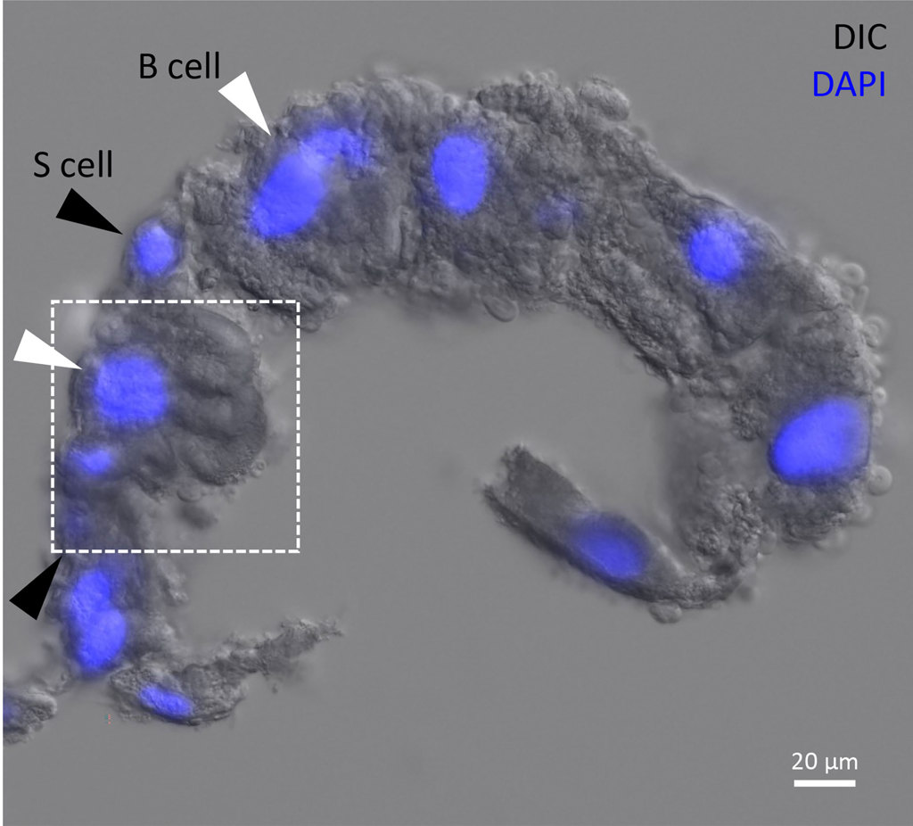
The fluorescent image of a hepatopancreas shows the cell nuclei labeled in blue. [Image: I. Pope, N.G.C. Ferreira, P. Kille, W. Langbein and P. Borri] [Enlarge image]
The researchers tested their scheme by feeding woodlice organic matter spiked with gold nanoparticles. Samples of the hepatopancreas were imaged with the FWM technique around four days later, clearly revealing the position of single gold nanoparticles within the multicellular organ. Other signals detected after longer pump–probe time delays were attributed to copper and iron structures that are known to accumulate inside the hepatopancreas.
This particular study exploited dissected samples, but in principle FWM microscopy could also be used to study living cells. Combined with its 3D imaging capabilities, the team believes that this relatively simple technique offers a valuable complement to synchrotron-based analysis. “Tracking gold nanoparticles within these organisms is the first step enabling further study to determine, for example, if gold is collected within specific cells, or if it can interfere with the metabolisms in high doses,” comments Langbein.