
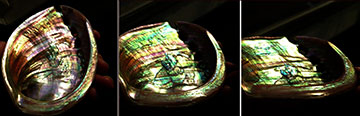
Nacre, or mother-of-pearl, is extremely fracture-resistant, which is one reason why researchers wanted to find a method to measure its thickness without harming it. [Image: Kats Lab / UW–Madison]
For centuries, mother-of-pearl—also known as nacre—has adorned precious objects like jewelry boxes and delicate buttons as a decorative element. The material lines the inside of some mollusk shells and is highly prized for its eye-catching iridescence and remarkable strength.
Probing the structure of nacre, however, remains challenging because existing methods rely on cutting the sample for analysis. Now, researchers at the University of Wisconsin–Madison, USA, have developed a new all-optical imaging technique to nondestructively characterize the structure of nacre and other layered biomaterials (PNAS, doi: 10.1073/pnas.2023623118).
A living record
Pupa Gilbert, professor of physics at the University of Wisconsin–Madison, has studied the architecture and possible formation mechanisms of nacre for more than a decade. She previously discovered that ancient nacre, which forms layer by layer like the rings of a tree, provides an accurate record of local ocean temperatures. Fossil samples up to 200 million years old reveal that thicker layers correspond to warmer years.
Such experiments require taking a cross-section of each shell and observing the layers with a scanning electron microscope. Samples are destroyed as a result, and it also proves difficult to probe large areas of nacre, which is typically not uniform across different areas.
Nondestructive optical imaging
Pupa Gilbert. [Image: UW–Madison]
At a seminar, Gilbert asked Mikhail Kats, associate professor of electrical & computer engineering at the University of Wisconsin–Madison, whether he could come up with an all-optical imaging technique that would not damage samples. In response, Kats and his colleagues developed hyperspectral interference tomography (HIT), a rapid and nondestructive way to spatially map the structure of nacre.
“Our technique is based on hyperspectral imaging, in which every pixel on an image contains a full reflectance spectrum in the visible and near-infrared,” said Kats. “We perform angle-dependent and polarization-dependent hyperspectral measurements of the shells, and then map the results to a computational model we built that predicts the reflectance spectra of possible nacre structures.”
From an optics perspective, nacre can be viewed as a disordered thin-film stack with defects, so they used thin-film modeling combined with a Monte Carlo technique to model the disorder. With HIT, the researchers found a previously unknown relationship between the age of the mollusk and the thickness of the aragonite tablets that make up nacre. Younger abalone grow thicker aragonite tablets compared with older abalone.
Future applications
HIT has several major advantages over existing techniques for the structural characterization of layered biomaterials in general. Aside from being nondestructive, the technique is very fast, inexpensive and portable. Kats and his colleagues look forward to more studies of nacre with Gilbert that may provide a more comprehensive view of climate history. They also believe that further investigation of its structure could inspire materials with greater toughness.
“Nacre has long been a subject of study in mechanics, because it is much more resistant to fracture than aragonite, which is the primary component,” said Kats. “So, it is important to try to further understand the formation and physical properties of nacre.”
