
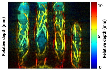
A U.S.-based research team has devised a biometric identification approach that leverages 3D images of finger veins to distinguish between different individuals. [Image: Y. Zhan et al., Appl. Opt., doi: 10.1364/AO.400550 (2020)]
The technique of photoacoustic tomography (PAT), an acousto-optical hybrid technique that combines the sensitivity of optical imaging with the penetration of ultrasound, can produce breathtakingly detailed images of cells and tissues for biomedical study. Now, a U.S.-based research team offers what could be a practical way to put PAT into service in another area: biometric scanning to verify people’s identity (Appl. Opt., doi: 10.1364/AO.400550).
The researchers have built a compact prototype scanner that collects photoacoustic data from a test subject’s fingers, pipes the data into a sophisticated modeling algorithm, and spits out a detailed 3D image of the pattern of blood vessels inside the finger. Such patterns—inherently unique to the individual, and buried beneath the skin—are effectively impossible to “spoof” or falsify. And, the researchers report, initial studies suggest that the new compact system they’ve developed can correctly tag an identity as much as 99% of the time using this internal fingerprint.
A light–sound one-two punch
PAT works by combining optical excitation with acoustic reporting (see “Multiscale Photoacoustic Tomography,” OPN, April 2018). A near-infrared, ns-to-ps laser pulse is fired into the tissue to be examined. When the pulse strikes a molecule in the tissue of interest—such as a vein—the molecule jumps to an excited state; as it relaxes, some of the energy re-emitted by the molecule is in the form of heat.
The sudden burst of heat creates a pressure wave inside the tissue that propagates at around 1500 m/s back out of the sample, and that can be read by an ultrasound transducer. The ultrasound signals resulting from repeated laser pulses are then sent to a computer for tomographic processing into a 3D image.
Because different biological tissues absorb at different optical wavelengths, PAT can provide unusual specificity—operators can target the tissue to be examined by selecting the right wavelength for the excitation beam. And because the scattering of acoustic waves in tissue is orders of magnitude weaker than optical scattering, the ultrasound delivery of the final signal can allow for far better spatial resolution at depth than in purely optical techniques.
From 2D to 3D
The idea of using finger vasculature as a potential biometric security tool has been broached before. But previous approaches have rested on purely ultrasound, optical or thermal techniques, and have provided only comparatively rough, 2D images.
Recognizing that 3D images would provide a far higher level of security and unambiguous identification, the research team—including scientists at the State University of New York, Buffalo, USA, and NEC Laboratories America, USA—turned to PAT. In a previous study published in 2018, several members of the team prototyped a photoacoustic-imaging platform based on palm scanning.
The prototype system provided results comparable to conventional biometric approaches. However, the authors of the new study note that it was “bulky and inconvenient,” and prone to deteriorating results given small errors in the subject’s hand placement.
Toward a compact setup
To get to something more practical, the team behind the new work focuses not on the palm vasculature, but on the veins in the fingers—and put some clever optical design to work.
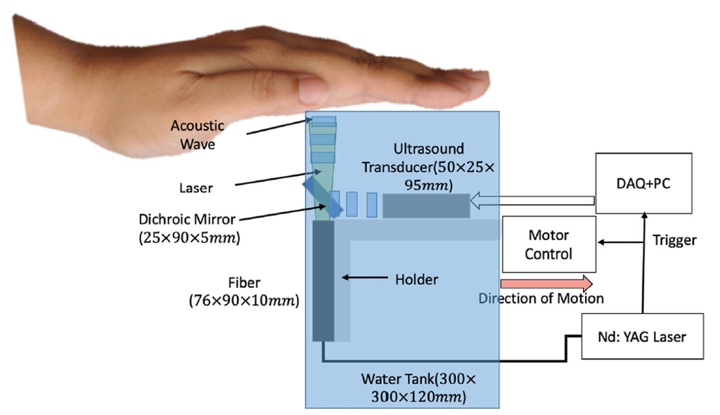
The use of a dichroic “cold” mirror that’s transparent to IR light but that reflects acoustic energy allowed a compact setup for the new system. [Image: Y. Zhan et al., Appl. Opt., doi: 10.1364/AO.400550 (2020)] [Enlarge image]
In the proof-of-concept of the new system—dubbed 3D Finger—the subject places his or her fingers on the top of a transparent water tank containing the light delivery system and a compact ultrasonic transducer. Light pulses from a 1064-nm Nd:YAG laser are delivered through fiber to a dichroic “cold” mirror that’s virtually transparent to IR light but that reflects more than 90% of incident acoustic energy.
After passing through the cold mirror, the laser light travels through the subject’s fingers, exciting the photoacoustic effect in the finger veins, and the resulting acoustic signal is sent back colinearly with the laser beam to bounce off the cold mirror. The sound waves then travel to the ultrasound transducer for recording and routing to a computer for imaging. The unit is mechanically scanned along the length of the subject’s hand to image the vasculature in the entire finger.
To put the finishing touches on the setup as a biometric-security system, the team also wrote an improved image-processing algorithm that filters out nonvascular features and noise to create a sharp 3D image of key vascular elements. A new matching algorithm, also built by the team, then creates a “vascular model” that can be numerically classified and compared with a reference scan of from the subject—to enable the real person to be distinguished from an impostor.
High accuracy
In tests involving 36 subjects, the research team found that the system provided an accuracy rate of greater than 99%. Not surprisingly, accuracy was particularly high when results from more than one of a given subject’s fingers were used in the comparison.
At this point the technique is not exactly rapid; each experiment took approximately 35 seconds. The researchers think that number can be hammered down, however. Indeed, they suggest that a scan time of as little as one second is conceivable through the use of LEDs or high-rep-rate laser diodes as the optical source, and by leveraging multichannel data acquisition cards for gathering the ultrasound signal.
The researchers are now working on these and other possible improvements to the system. They are even looking, eventually, at porting the system to smartphones, some of which, the authors note, already include ultrasound capabilities.
In a press release accompanying the research, Giovanni Milione, a researcher at NEC Laboratories, sketched out some of the prospects the team is exploring. “We envision this technique being used in critical facilities, such as banks and military bases, that require a high level of security,” he said. “With further miniaturization, 3D vein authentication could also be used in personal electronics or be combined with 2D fingerprints for two-factor authentication.”
