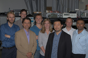
Co-authors of the "imaging flow analyzer" paper (left to right): Dino Di Carlo, Daniel R. Gossett, Bahram Jalali, Coleman Murray, Nora Brackbill, Keisuke Goda, Jost Adam and Cejo K. Lonappan. Photo courtesy of K. Goda.
Finding a single cancer cell circulating among billions of blood cells is a difficult task, but a new device that performs ultrafast imaging and classification of moving particles could make the job easier.
Researchers at the University of California at Los Angeles (UCLA, U.S.A.) developed an “imaging flow analyzer” that takes blur-free pictures of fast-moving microparticles, then sorts out the images (Proc. Natl. Acad. Sci. USA, doi: 10.1073/pnas.1204718109). The device can examine 100,000 blood cells per second, far more than conventional microscopy.
Many cancer patients die from their disease’s metastasis, not from the primary tumor, according to the paper’s lead author, Keisuke Goda. Conventional optical microscopes are not designed to study these so-called circulating tumor cells in the bloodstream because they are extremely rare compared with normal cells. Finding these unusual cells in a patient’s blood sample requires the rapid analysis of a huge number of cells.
The flow analyzer incorporates a unique camera invented by OSA Fellow Bahram Jalali’s group at UCLA three years ago (OPN, July/August 2009, p. 9). Dubbed serial time-encoded amplified microscopy, or STEAM, the camera encodes 2-D image data into broadband near-infrared laser pulses, optically amplifies the pulses and stretches them in time.
The device routes a sample’s blood cells through a sinuous microfluidic channel, where inertial forces focus them into a single-particle stream by the time they pass through the STEAM camera’s light pulses. A customized high-speed field-programmable gate array (FPGA) processes the incoming images continuously and starts the image-sorting process, completed in an additional CPU. The system avoids the usual bottlenecks in the data-transfer process, says Goda, the program manager of Jalali’s photonics group.
To test the proof-of-principle device, the UCLA group used a blood sample containing a small number of breast cancer cells, which are roughly twice as big as other blood cells. At a flow speed of 4 m/s, the analyzer screened 100 million cells and found 75 percent of the cancerous ones.
Next, Goda and his colleagues will work on improving the device’s image quality and refining the signal processing algorithms while they seek opportunities for clinical studies.
