
 An Egyptian sarcophagus with a mummy inside. [banusevim / Getty Images]
Spectroscopy plays a crucial role in the field of cultural heritage. It can assist with diagnostics, analysis and characterization of a cultural object’s material composition, guide the development and monitoring of new conservation techniques, or identify the optimal environmental conditions for preventive conservation. Unsurprisingly, several studies have been published in recent years exploring the application of diverse spectroscopic methods in this field.
However, analyzing rare historical or archaeological samples requires special considerations and can pose great challenges to spectroscopists. The objects are fragile and irreplaceable, ideally requiring noninvasive and nondestructive techniques. Furthermore, many have undergone aging, degradation or interactions with environmental factors that can alter their original chemical composition and might complicate their identification. In addition, archeological or historical samples typically have complex chemical compositions that challenge standard analysis approaches.
Spectroscopic techniques need to be user friendly and provide results that are clear and easy to understand by non-technically-trained people.
In addition to sensitivities that come with how precious the samples are, there are also various demands with regard to the measuring instruments and system engineering. For a broad and successful application outside of laboratories, measurement instruments need to be fast, robust, affordable, reliable and easy to operate. Moreover, the techniques need to be user friendly and provide results that are clear and easy to understand by non-technically-trained people. It should also allow for mobile analysis so that it can be utilized to examine objects in fixed locations.
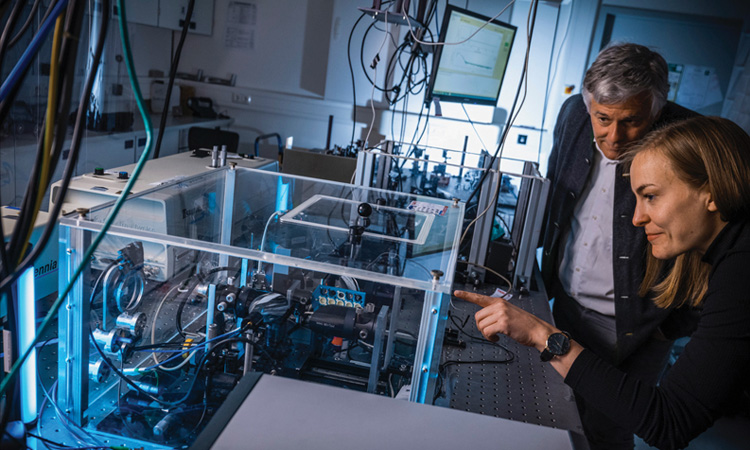 Martin Koch (back) and Lara Heidrich (front) in the lab in front of a setup used for terahertz time-domain spectroscopy of natural resins. [J. Hosan]
Spectroscopic methods: Strengths and limitations
All spectroscopic methods offer advantages and disadvantages. When selecting the most suitable technique(s) for thorough material classification, several application-dependent aspects should be considered.
Raman spectroscopy
Raman spectroscopy is one of the most appreciated analytical techniques in the field of cultural heritage science due to several factors, such as the relatively quick time of analysis as well as its ability to identify both inorganic and organic molecules. Using this technique, the sample is illuminated with monochromatic light, typically from a laser, and inelastically scattered light is detected. The energy shift between the incident and emitted light is called the Raman shift and is based on the excitation of, for instance, molecular vibrations or phonons. It provides a unique spectral fingerprint of the sample, yielding information about molecular bonding, crystallinity, phase transitions and chemical composition. Portable or handheld systems are available, and several in-situ mobile Raman applications on cultural heritage objects have been reported, such as the identification of rock art painting materials or the investigation of medieval manuscripts and paintings. However, local heating or photodegradation of the sample may occur, and the most serious problem with Raman spectroscopy is the possible occurrence of fluorescence, which can interfere with the measurement. Fluorescence involves the absorption of incident light and the subsequent excitation of a molecule to a higher electronic energy level. From this excited state, the molecule relaxes to the ground state via vibrational and radiative transitions—the latter being much more intense than those observed in Raman scattering. Therefore, the spectral window in which fluorescence excitation occurs should be avoided when selecting the laser excitation wavelength.
Laser-induced breakdown spectroscopy
Laser-induced breakdown spectroscopy (LIBS) is a specific type of atomic emission spectroscopy. During LIBS, a high-power pulsed laser beam is focused onto the surface of the material of interest. Consequently, a small portion of the material is ablated and a hot plasma containing free electrons and ionized atoms is created. Upon relaxation of the plasma, distinct emission lines characteristic of the elements present in the ablated matter can be detected. For instance, LIBS has been used for analytical assessment and chemical mapping of the façade of the Malaga cathedral in Spain. A major advantage of this method is that it is very fast and does not require sample preparation. However, it is a minimally invasive method and can lead to partial destruction of the sample, limiting its use for highly valuable objects.
Nuclear magnetic resonance spectroscopy
Nuclear magnetic resonance (NMR) spectroscopy is based on the fact that the resonance behavior of atomic nuclei with magnetic moments under electromagnetic irradiation is affected by tiny differences in magnetic field shielding caused by the surrounding atoms—and is mostly influenced by the atoms that are covalently bound to that specific atom. Hence, NMR spectroscopy can provide comprehensive information about the chemical structure of a material, as the NMR spectra obtained are unique to functional groups or individual compounds. Applications in the field of cultural heritage include, for instance, assessing the impact of aging on materials, monitoring conservation and restoration efforts, and completing stratigraphic analysis in paintings and frescoes. However, NMR analysis is characterized by its time-consuming nature, high acquisition and operating costs, and complex data interpretation. Recently, the use of artificial intelligence (AI) tools has assisted spectroscopists in the interpretation of NMR data and offers potential advantages, particularly in the analysis of natural products.
Fourier-transform infrared spectroscopy
Fourier-transform infrared (FTIR) spectroscopy works predominantly in the mid-infrared. It is a well-established technique—the gold standard for broadband vibrational spectroscopy. With IR radiation, intramolecular vibrational modes can be excited, yielding information via absorption bands corresponding to certain functional groups present in the molecules or through molecular fingerprints characteristic for specific compounds. Extensive databases are available as well as portable or handheld systems. A well-known example of its use is the analysis of oil paintings, as these consist of several layers containing a variety of organic and inorganic components. In addition, IR spectroscopy—such as Raman spectroscopy—can be used to estimate sample age and assess microstructural changes. When materials undergo weathering, their crystalline structures can become disrupted, leading to increased amorphization. This structural disorder affects the vibrational modes of molecules, resulting in broader and sometimes shifted spectral peaks. However, a planar surface or low contact pressure is required for sampling, which may make it unsuitable for the analysis of fragile samples.
Terahertz spectroscopy
Longer wavelengths are used in terahertz (THz) spectroscopy. THz time-domain spectroscopy (TDS)—the most commonly used spectroscopy method in this frequency range—probes intramolecular vibrations of larger groups of atoms or vibrations of entire molecules against each other, such as intermolecular or phonon-type vibrations in a molecular crystal. This method yields limited spectral detail for non-crystalline samples or if the molecules are too large (for example, proteins). Instead, it provides a continuous absorption spectrum, which is not characteristic and therefore of little significance. However, even though the spectral information obtained from THz TDS may be limited for complex mixtures, the technique offers unique advantages and possibilities in the context of cultural heritage. First, THz radiation can penetrate through many optically opaque materials, such as paper, fabric, paint, wood or ceramics, allowing for the sub-surface analysis of various objects. Second, in contrast to computed tomography scans, non-ionizing radiation is used, which simplifies the analysis of objects as their safety and that of the operator of the device is not jeopardized. Third, due to the pulsed nature of the technique, it can be used to reconstruct cross-sectional images of the internal structure of objects. These stratigraphic images are particularly useful for identifying sub-surface air gaps, monitoring the consolidation process of a painting or determining cracks and paint layer separation on artwork.
Photoluminescence spectroscopy
Using photoluminescence (PL) spectroscopy, the object under investigation is irradiated by a laser or strong LED with a fixed wavelength. This leads to the excitation of electronic transitions in the material and subsequent relaxation and emission of photoluminescent light. For solids, the emitted light does not consist of sharp emission lines but is rather a continuous and broad emission spectrum. However, PL spectra from different substances differ slightly, allowing for material identification and characterization.
A related method is photoluminescence excitation (PLE) spectroscopy, where the PL emission is detected at a fixed wavelength and the excitation wavelength of the light source is varied. The big advantages are that simple and low-cost devices are available and that the samples can be arbitrarily shaped. PL spectroscopy represents a noninvasive and nondestructive technique, which is of great importance when investigating precious samples. The use of PL spectroscopy in cultural heritage material studies has been limited to date due to the rather broad and uncharacteristic emission spectra, which may not be able to provide clear evidence of the chemical identity of materials. Nonetheless, even broad characteristics might be helpful to distinguish or classify materials. In combination with affordable and versatile sampling devices and the ongoing development of meaningful databases, it represents a promising tool in the context of cultural heritage studies.
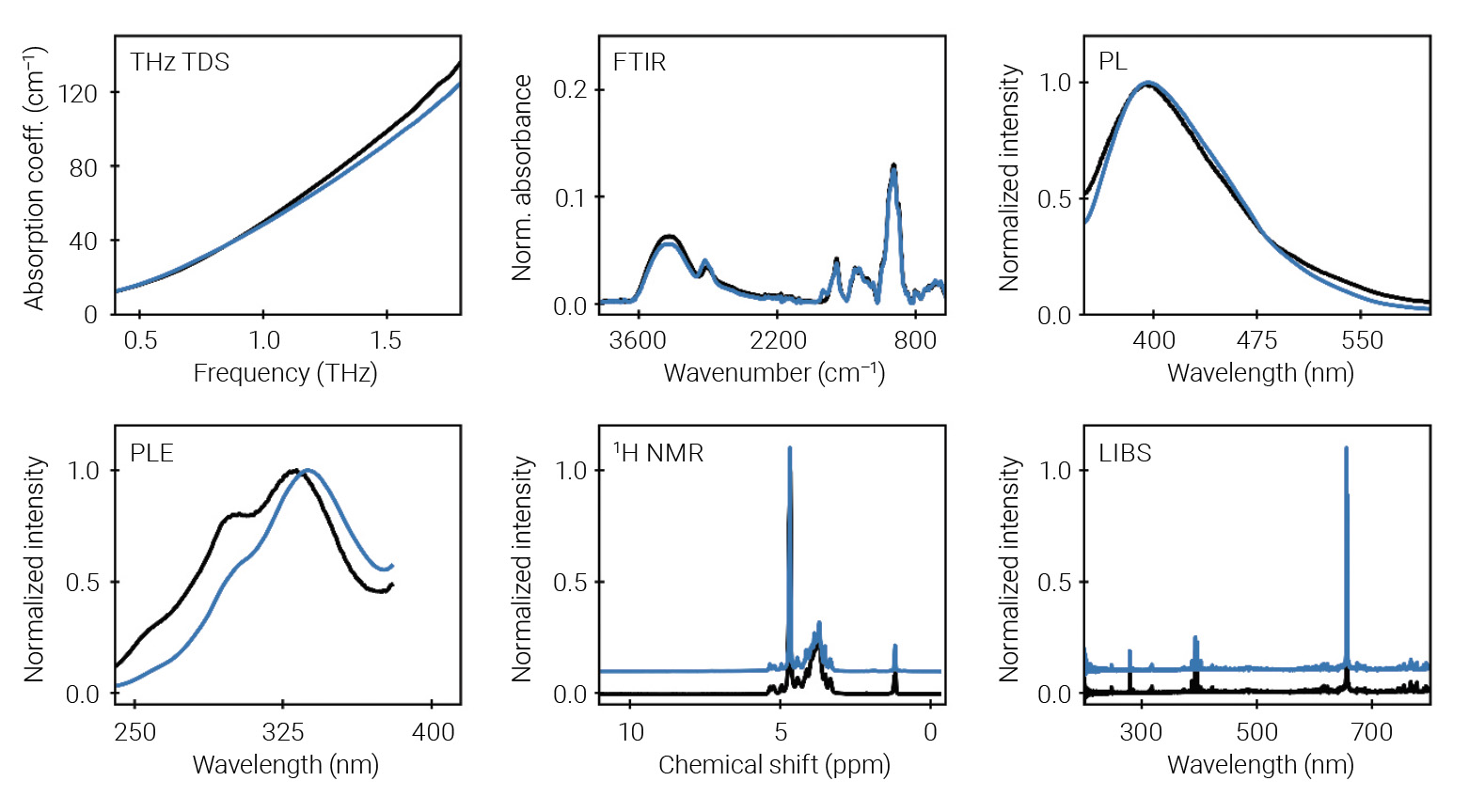 [Enlarge image]Comparison of the results obtained by the different spectroscopic techniques for gum arabic (black lines) and an unknown sample from an Egyptian market (blue lines). The spectra indicate that the market sample is probably also gum arabic. For clarity, the 1H NMR and LIBS data is plotted with an offset of +0.1 (blue spectra). Please refer to the original publication for a detailed discussion of the results presented. [Heidrich et al. “Multispectral investigation of natural resins,” Archiv der Pharmazie 357, 11, e2400517 (2024).]
Secret mummification recipes
Egyptian mummies have fascinated scholars for centuries. Yet the exact recipes of the balms used for mummification remain uncertain, as there are only a few ancient Egyptian texts mentioning the exact ingredients.
The exact recipes of the balms used for mummification remain uncertain, as there are only a few ancient Egyptian texts mentioning the exact ingredients.
The use of molecular analyses to help in identifying the ingredients of ancient Egyptian embalming materials has been of great interest in recent decades. Traditionally, gas chromatography–mass spectrometry or liquid chromatography have been used to identify the base components of the embalming materials—typically natural resins. However, both techniques are destructive due to the required use of organic solvents and do not allow the analysis of valuable archaeological samples. This raises the question of whether the use of spectroscopic methods can provide a methodological advantage in resin residue analysis.
Besides mummification, natural resins have also been used as protective varnishes and dyes in ancient painting techniques or as remedies in a medical context. Hence, the successful spectroscopic analysis of natural resins can be useful not only in the field of Egyptology, but also in medicine and art history.
Investigating natural resins
Recently, we explored and compared the use of five spectroscopic techniques as alternative methods to the established analytical procedures for natural resin analysis. In the course of this, we used THz TDS, LIBS, PL/PLE, NMR and FTIR spectroscopy. In total, three unknown samples from an Egyptian market were studied alongside 15 resin samples of known origin. The latter included frankincense, gum arabic, myrrh, elemi, mastic and dammar, which have been suggested in recent studies to be part of the embalming mixtures.
LIBS and THz TDS provided limited information for resin characterization, and classification is only possible according to the major structural class of the resins (for instance, triterpenic or gum resin). However, this is not particularly surprising given the structural information that can be obtained from both techniques. Natural resins are complex mixtures of more than 100 different substances. Hence, no coherent long-range motions can be excited by THz radiation—as would be the case for molecular crystals—leading to uncharacteristic THz spectra. E. Stübling and colleagues at Philipps-Universität Marburg, Germany, and the University of Zürich, Switzerland, reported that it was possible to nondestructively identify embalming materials in two unknown bandages by further analyzing THz TDS data through principal components analysis. Using LIBS, structural information about the elemental composition of a material can be obtained. As the resins differ mainly due to their different organic ingredients, which in turn have similar elemental composition—carbon, hydrogen and oxygen—only minor differences in the LIBS data can be expected.
In contrast, it is possible to classify the resin samples more precisely using FTIR, NMR and PL/PLE spectroscopy. Furthermore, by linking NMR analysis with AI, further sub-classification of the individual resins can be achieved. However, the application of both FTIR and NMR analysis will be limited due to the partial destruction of the sample, the complex analysis of the obtained data and the expensive equipment required. Although the results obtained by PL/PLE spectroscopy are not as informative as the NMR data, the PL/PLE spectra of the various resins differ considerably, enabling a fast, affordable and nondestructive material classification.
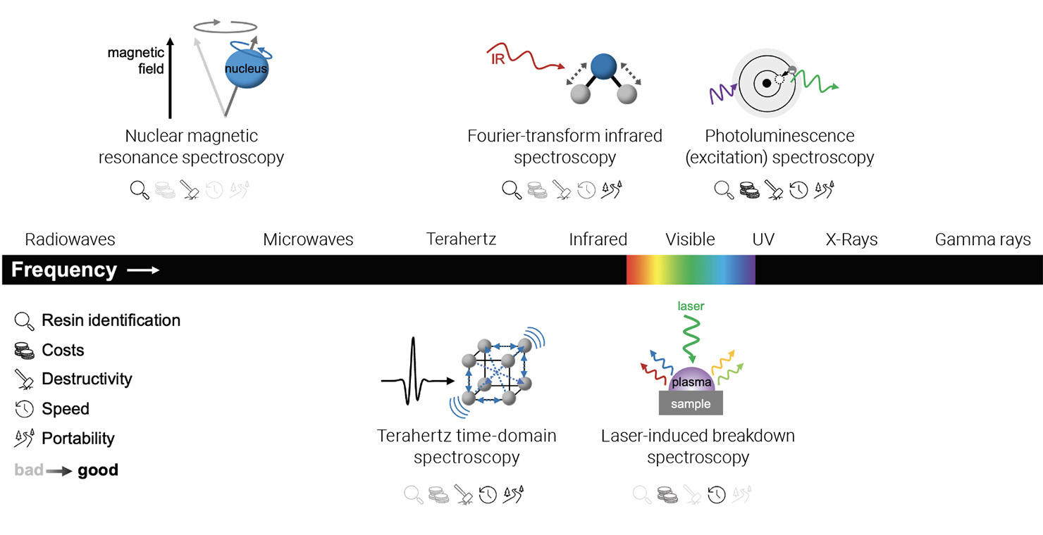 [Enlarge image]Schematic electromagnetic spectrum (unscaled) with spectroscopic techniques used for natural resin identification as well as the limits and strengths of the techniques. [L. Heidrich]
Looking beneath the surface
THz spectroscopy is not only suitable for obtaining spectral information about an object but also allows the near-surface structure of objects to be clarified. This can be achieved using THz tomography, which was first demonstrated by Daniel Mittleman and colleagues at Bell Laboratories in 1996. The pulses used in this measurement method are reflected from any interface between materials with different dielectric properties just like in optical coherence spectroscopy, which is a standard method in the near-infrared frequency range. Since most materials show a certain residual absorption and are rough and therefore scatter, the penetration depth is usually limited to a few millimeters.
Initially, only flat samples, which were moved through the focus in a raster pattern, could be examined. However, fiber-coupled THz spectrometers, which have been available for many years, allow greater flexibility. This means that a THz measuring head can be attached to a robot arm and moved flexibly, allowing examination of objects with curved surfaces. The group at Philipps-Universität Marburg, for example, used it to examine a 3,000-year-old mummified hand.
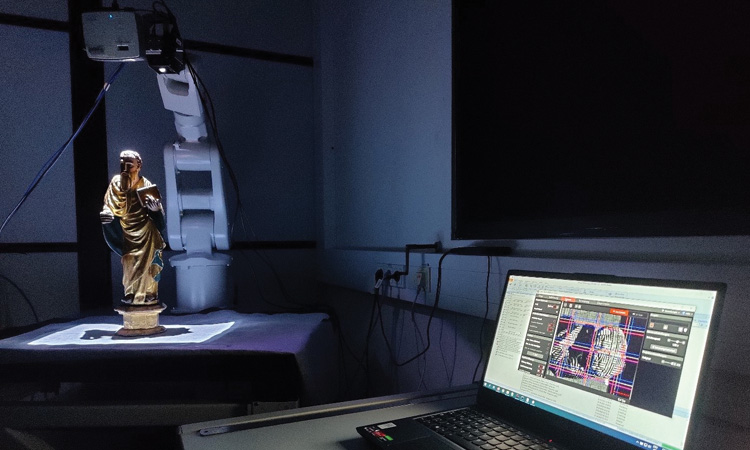 Robotic-based THz imaging system used for the investigation of a sculpture from the right wing of the Trinity Altar, Hildesheim/Roemer- and Pelizaeus-Museum, Hildesheim, Germany. [K. Krügener, University of Applied Sciences and Arts (HAWK) Hildesheim, Göttingen, Germany]
Luminescence of modern environmental samples
As mentioned earlier, PL spectroscopy is a fast method for obtaining initial information about natural resins. Yet, not only resins but most substances, with the exception of metals, show a weak luminescence when optically excited. This property can be used in the field of cultural heritage and in other areas.
One example is the cost-effective identification of microplastics. PL spectroscopy allows plastic particles to be distinguished from other materials, for example. However, it is also possible to classify the different plastics from one another to a certain degree. This also works with relatively inexpensive lasers around 405 nm. The luminescence intensity and the discrimination accuracy generally increase when a shorter excitation wavelength is selected.
Luminescence spectroscopy can be an inexpensive tool to collect information about objects in the field, which is of great importance in order to understand the effects of microplastics.
Overall, luminescence spectroscopy can be an inexpensive tool to collect information about objects in the field, which is of great importance in order to understand the effects of microplastics on living organisms as well as their distribution and composition in the environment.
Future directions
It is desirable to analyze cultural heritage objects on-site using multiple nondestructive, complementary spectroscopic methods simultaneously. Such an approach would provide a wide range of results, mutually compensating for the limitations of the individual techniques and leading to unique findings about the objects under examination. In any case, even when the chemical composition of a material or object can be determined using spectroscopy, understanding its original function requires collaborations among scientists, historians and conservators to interpret findings within their cultural and historical contexts. Addressing these challenges necessitates continuous advancements in analytical techniques, development of extensive reference databases, and interdisciplinary collaborations to gain deeper insights into cultural heritage objects.
Lara Heidrich and Martin Koch are with the Department of Physics at the Philipps-University of Marburg, Germany.
References and Resources
-
G. Bitossi et al. “Spectroscopic techniques in cultural heritage conservation: A survey,” Appl. Spectrosc. Rev. 40, 187 (2005).
-
P. Vandenabeele and M.K. Donais. “Mobile spectroscopic instrumentation in archaeometry research,” Appl. Spectrosc. 70, 27 (2016).
-
E. Stübling et al. “On the potential of THz time-domain spectroscopy to identify typical ancient Egyptian embalming materials,” J. Infrared Millim. Te. 40, 763 (2019).
-
E. Stübling et al. “Application of a robotic THz imaging system for sub-surface analysis of ancient human remains,” Sci. Rep. 9, 3390 (2019).
-
J. Ornik et al. “Could photoluminescence spectroscopy be an alternative technique for the detection of microplastics? First experiments using a 405 nm laser for excitation,” Appl. Phys. B 126, 15 (2020).
-
K. Peiponen et al. “Detecting microplastics with optics,” Opt. Photon. News 31(11), 24 (November 2020).
-
B. Lotter et al. “Identifying plastics with photoluminescence spectroscopy and machine learning,” Sci. Rep. 12, 18840 (2022).
-
M. Koch et al. “Terahertz time-domain spectroscopy,” Nat. Rev. Methods Primers 3, 48 (2023).
-
L. Heidrich et al. “Multispectral investigation of natural resins,” Arch. Pharm. 357, 11, e2400517 (2024).
For additional references and resources, visit: optica-opn.org/link/0725-spectroscopy.
