
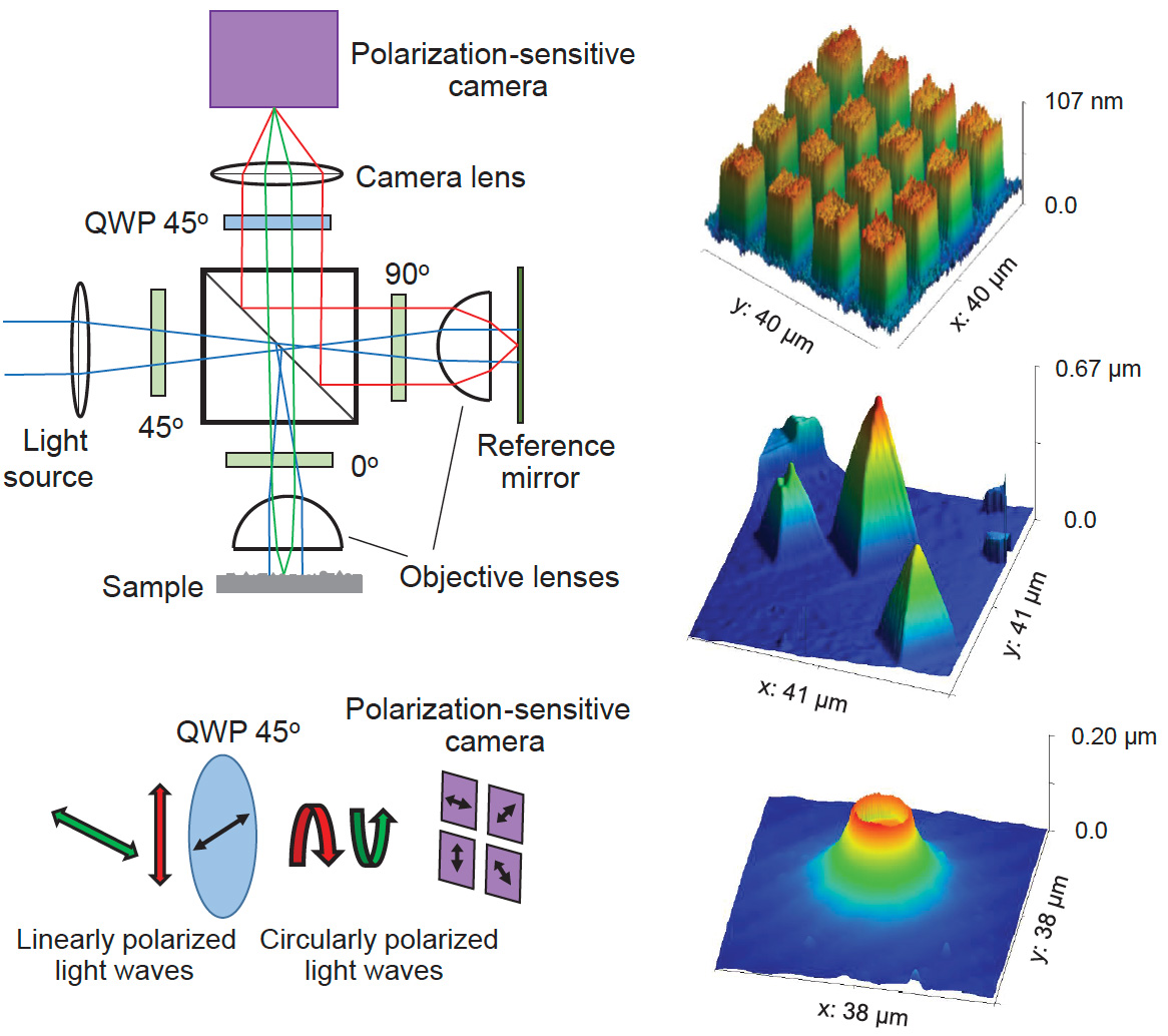
 [Enlarge image]Left: Schematic of the single-shot wide-field interference microscope based on a Linnik interferometer (top). Illustration of the transformation of orthogonally polarized light into oppositely handed circular polarizations after the quarter-wave plate (bottom). Right: Examples of 3D surface reconstructions obtained from single-shot interference patterns: 100 nm atomic force microscope height standard (top), chemical vapor deposition diamond surface (center) and red blood cell (bottom). Image acquisition time 3 ms.
Unlike most optical instruments that form images based on light intensity, interferometric techniques extract information from the phase of light. This phase sensitivity enables spatial resolution beyond the diffraction limit. Among the various interferometer configurations available,1,2 the Linnik interferometer3-5 stands out as a particularly promising platform for microscopy applications.
We present a wide-field microscope based on the Linnik interferometer that employs an incoherent light source (λ₀ = 470 nm, ∆λ = 30 nm). Light passes through a non-polarizing beam splitter and is directed toward the sample and reference arms, each with an Olympus UP-LFLN 60× objective. The reflected beams recombine at the detector, where a polarization-sensitive camera records four polarization states simultaneously. The setup ensures that reflections from the sample and reference mirror are linearly polarized in orthogonal directions. After passing through a quarter-wave plate, they are converted into oppositely handed circular polarizations. The camera then captures four interference images with π/2 phase shifts,4,5 enabling complete phase reconstruction for 3D imaging.
A polarizer at 45° before the beam splitter is essential,5 since interference only occurs when photon paths through the interferometer are indistinguishable, analogous to Young’s double-slit experiment.
Image acquisition requires only a few milliseconds, depending on sample reflectivity and camera sensitivity. This short exposure makes the system highly resistant to vibrations and drift. The microscope achieves 10 nm vertical (Z) resolution, while lateral (X–Y) resolution remains diffraction limited.
We also implemented an alternative configuration, replacing the 0°/90° polarizers with quarter-wave plates and adding a polarizing beam splitter, similar to prior Fizeau interferometer work. This simplifies alignment and allows brightness control between the arms. However, the extinction ratio of a polarizing beam splitter is generally lower than that of high-quality polarizers, which can reduce interference contrast.
In summary, the single-shot, wide-field Linnik microscope enables rapid topographic imaging with nanometer-scale vertical resolution. Its fast, stable operation makes it well suited for video-rate imaging of dynamic processes. The use of an incoherent source offers wavelength flexibility and keeps costs low, broadening applicability.
Researchers
Sergei V. Anishchick and Marcos Dantus, Michigan State University, USA
References
1. D. Malacara et al. Interferogram Analysis For Optical Testing, Taylor & Francis, CRC (2005).
2. D. Malacara (Ed.) Optical Shop Testing, Wiley (2007).
3. W.P. Linnik, Proc. Acad. Sci. USSR, 1, 208 (1933).
4. S.V. Anishchik and M. Dantus, J. Optics, 26, 115602 (2024).
5. S.V. Anishchik et al. Proc. SPIE, 13325, 1332509 (2025).
