
Scatterings
Stimulated Raman Scattering Improves Video of Living Tissue
Researchers have sped up the stimulated Raman spectroscopy imaging process to make real-time imaging of surface tissue cells practical.
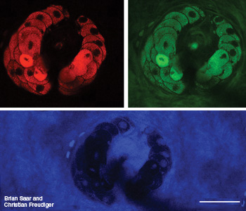 Label-free tissue imaging of a sebaceous gland wrapping around a hair in the viable epidermis of mouse skin. Image in red shows lipid-rich gland cells; green image shows new protein-rich structures such as a hair in the center; blue shows inverse image contrast from the gland.
Still images work for some biomedical applications, but video provides scientists with real-time imaging of drug diffusion in tissues, among other uses. Researchers have now speeded the stimulated Raman spectroscopy (SRS) imaging process to make real-time imaging of surface tissue cells practical (Science 330, 1368).
…Log in or become a member to view the full text of this article.
This article may be available for purchase via the search at Optica Publishing Group.
Optica Members get the full text of Optics & Photonics News, plus a variety of other member benefits.
