
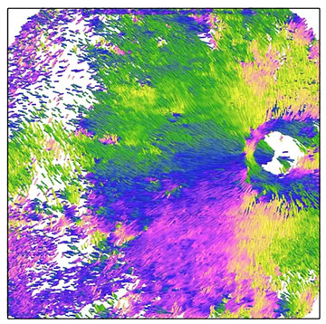
Researchers successfully applied PS-OCT to assess various properties of the collagen fibers that make up the sclera, the outer white of the eye. [Image: Department of Ophthalmology and Visual Science, TMDU]
The sclera, or white part of the eye, acts as a protective outer covering that also helps maintain the eye’s shape. The thickness of the sclera can be imaged with traditional optical coherence tomography (OCT), but observing the details of the collagen fibers that make up the tissue remains a challenge.
Now, researchers in Japan have found that the addition of polarization-sensitive OCT (PS-OCT), which uses the polarization of light as the image contrast mechanism, can reveal the density and orientation of collagen fibers in the inner and outer layers of the sclera (JAMA Ophthalmol., doi: 10.1001/jamaophthalmol.2024.0002). The approach may contribute to a better understanding of—and possible treatments for—scleral pathologies that affect vision.
Visualizing collagen fibers
A dome-shaped macula in highly myopic, or nearsighted, eyes can lead to gradual mild-to-moderate vision loss over a few years. The macula, the part of the retina located at the back of the eyeball, is essential for central vision. An inward protrusion of the macula may result from a thickening of the macular sclera.
“The sclera, composed of collagen fibers, plays an important role in protecting the retina, optic nerve and other nerve tissues in the eye,” said study author Kyoko Ohno-Matsui, Tokyo Medical and Dental University, in a press release accompanying the results. “Therefore, abnormalities in the shape of the sclera can cause various complications leading to blindness.”
Ohno-Matsui and her colleagues wanted to go beyond measuring thickness and examine the collagen fibers in the sclera of living human patients, which had never been done before. They constructed a prototype PS-OCT system to visualize the scleral collagen fibers in highly myopic eyes with and without dome-shaped macula.
Ohno-Matsui and her colleagues wanted to go beyond measuring thickness and examine the collagen fibers in the sclera of living human patients, which had never been done before.
Toward new therapies
Because the sclera has a polarization property called birefringence, in which the refractive index depends on the state of polarization, the PS-OCT system could determine the axis of orientation or the optic axis of the birefringence that is related to the orientation of the fiber bundles.
The PS-OCT instrument employed a tunable vertical-cavity surface-emitting laser with a center wavelength of 1050 nm, a bandwidth of 100 nm and a repetition rate of 100 kHz. The interferometer was configured to measure all elements of the Jones matrix of the sample in a parallel manner. The researchers measured 89 eyes from a total of 72 patients, mostly adults over 50 years old.
PS-OCT images revealed low-density, radially oriented fibers in the inner layer and high-density, vertically oriented fibers in the outer layer of the sclera. Patients with dome-shaped macula had aggregated and thickened collagen fibers in the inner layer of the sclera, whereas those in the outer layer were compressed and thinned.
“Given the common occurrence of scleral pathologies, such as [dome-shaped macula] and staphylomas in eyes with myopia, recognizing fiber patterns could provide important insights that may be relevant to developing targeted therapies to address scleral abnormalities early and mitigate potential damage to the overlying neural tissue,” said study author Masahiro Yamanari, in a press release accompanying the research.