
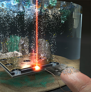
Artist’s impression of the field-induced active collection of nanoplastics using a synergetic combination of an electrophotonic tweezer and surface-enhanced Raman scattering. [Image: Korea Institute of Science and Technology]
Microplastics are now found everywhere on our planet—even in freshly fallen snow in Antarctica. Though they are still being studied, these plastic particles could pose significant threats to Earth’s ecosystem and human health. For example, when the particles disintegrate and shrink to less than 100 nm, they could invade various parts of human body through inhalation, ingestion, absorption through the skin or even penetration through cell membranes.
In their latest study, researchers in the Republic of Korea report combining an electric “tweezer” with plasmonic nanorods to develop a real-time separation and detection system for nanoplastics (plastic particles smaller than 1 μm) (ACS Nano, doi: 10.1021/acsnano.2c07933). Using a single platform, they say they can catch, separate and analyze the plastic particles within seconds, significantly reducing the time required for this process, which was previously at least one day.
Electric tweezer
Nanoplastics are generally hard to detect because of their small sizes and low concentrations in the environment. Raman spectroscopy has become one of the most promising techniques for analyzing nanoplastics, thanks to the specificity of a molecule’s vibrational frequency and its ability to spot the particles in situ.
However, find nanoplastics with Raman spectroscopy, the target particles need to be located within a micrometer-scale laser spot. This is hard to achieve because the Brownian random walk is the only mechanism driving the fragments around. Scientists have suggested pretreatment techniques for gathering nanoparticles, but the methods are cumbersome and time-consuming. In addition, the Raman scattering cross-sections of nanoparticles are quite small, leading to low signal-to-noise ratio.
To improve the use of Raman spectroscopy, a team led by Yong-Sang Ryu at the Korea Institute of Science and Technology combined two phenomena: dielectrophoresis (DEP), where a non-uniform electric field exerts a force on dielectric particles, and AC electro-osmosis (ACEO), where AC voltages on microelectrode structures generate flow of fluids. The result was the DEP-ACEO-Raman tweezer (DART)—an electric tweezer.
Amplifying with gold nanorods
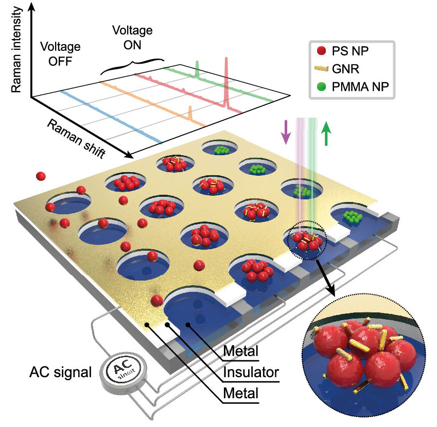
Schematic diagram of the DEP-ACEO-Raman tweezer (DART) system for real-time on-site Raman detection of nanoplastics. [Image: E.-S. Yu et al., ACS Nano, doi: 10.1021/acsnano.2c07933 (2023)] [Enlarge image]
For their experiment, the researchers created the DART using three layers: a top electrode layer made of gold; an insulating layer made of polydimethylsiloxane (PDMS); and a bottom electrode layer made of indium tin oxide (ITO). Both the top and middle layers were patterned with 2D arrays of 10-µm-diameter holes, repeating every 30 µm.
After placing their device at the bottom of a solution containing commercially available polystyrene particles, the researchers applied AC across the electrodes. The electric current instantly caused the plastics to densely gather into the center of the holes, improving the chances of detecting the particles using Raman scattering. Further tests revealed that the system is capable of detecting 200-nm nanoplastics even at a few mg/L when 2 V of AC was applied with the frequency of 10 kHz.
The researchers then took an additional step to improve the detection rate, as nanoplastics tend to exist in the ecosystem in even lower concentrations, and the DART system alone has difficulties detecting particles smaller than 100 nm. When they added gold nanorods to the sample, the nanorods concentrated in the center of the tweezers’ holes along with the nanoplastics, creating surface-enhanced Raman scattering hotspots. The researchers estimate that, with 5 V applied, the detection limit for 30-nm polystyrene particles should be about 1 µg/L.
Real-world application
The researchers write that, to best of their knowledge, the DART method demonstrates the highest detection performance so far for an in situ detection technique that doesn’t require any pretreatment—given that the nanoplastic concentration and the particle size they measured were both the lowest (10 µg/L) and the smallest (30 nm) ever detected. And because the real-world nanoplastic concentration is about 20 µg/L in seawater, they say their method’s potential for on-site application is evident.
The team also writes that its system can be applied to detect other types of targets, such as composite materials, nonpolymeric nanomaterials and even bioparticles such as exosomes and viruses.
Ryu now works at Korea University.
