

An international group of researchers has shown how a ytterbium-based soft X-ray laser can probe the sub-picosecond dynamics of ferrimagnets made from rare-earth elements. [Image: Ella Maru Studio]
An international group of scientists has used a table-top X-ray laser to probe the exceptionally swift magnetic dynamics of a rare-earth-element-based ferrimagnet—a type of material particularly well-suited to spintronic devices (Optica, doi: 10.1364/OPTICA.443440). The researchers say that their demonstration shows the potential of relatively small and cheap laser sources for visualizing and manipulating magnetic nanostructures as well as studying fleeting electronic motion in biological systems.
Scrutinizing rare-earth elements
Spintronics offers the potential of a radical alternative to conventional electronics, thanks to its exploitation of electrons’ intrinsic angular momentum, or “spin,” as well as charge. The fact that spins can be flipped on far shorter timescales than are needed to shunt charge around a circuit suggests that spintronic-based processors and memories might be quicker and less energy-intensive than today’s microelectronic circuits.
Among the most promising materials for fabricating spintronic devices are multi-component nanostructured systems based on rare-earth elements. These have particularly small features and are capable of being driven efficiently, but as with any candidate material, their magnetic behavior must be scrutinized in great detail. Scientists have, to date, studied a number of rare-earth elements alloyed to transition metals but have done so using short wavelength X-rays from large, centralized facilities such as synchrotrons and free-electron lasers.
“Soft” X-rays
In the latest work, Tadas Balciunas at the Technical University of Vienna in Austria and colleagues in Europe, Canada and China instead show the potential of smaller lasers exploiting high harmonic generation. This non-linear process involves an intense laser pulse being fired into a solid, liquid or gas target and generating a series of harmonics that can have frequencies many orders of magnitude higher than that of the source beam.
Laser systems that use this technique to generate “soft” X-rays at relatively long wavelengths can be used to carry out what is known as X-ray resonant magnetic scattering. By preferentially scattering X-rays whose energies match the absorption edges of the elements within a sample, this tool provides both diffraction and spectroscopic information on the material in question—a combination not possible with conventional microscopy or scattering studies.
Compared with straightforward spectroscopy, however, resonant magnetic scattering suffers from low scattering cross-sections and so requires a very intense source of photons. It also leads to a merging of absorption edges when being used to study multi-component systems at long X-ray wavelengths. The trick is to raise photon energies just enough that they cover the absorption edge of 4d electrons in rare-earth ferrimagnets, but lasers based on optical parametric amplifiers can only do so by sacrificing efficiency.
Targeting the 4d edge
Balciunas and colleagues overcome this problem by instead turning to ytterbium-based laser technology, which in recent years has shown its potential for scaling to high powers while remaining compact. The researchers hooked up an infrared ytterbium laser to a hollow-core fiber in order to compress its pulses and then sent those pulses through a gas cell to generate the high harmonics. By optimizing the laser wavelength as well as the type and pressure of the gas (they found that helium was best), they were able to generate very bright X-rays with energies of up to 220 eV and pulse lengths of just 25 fs.
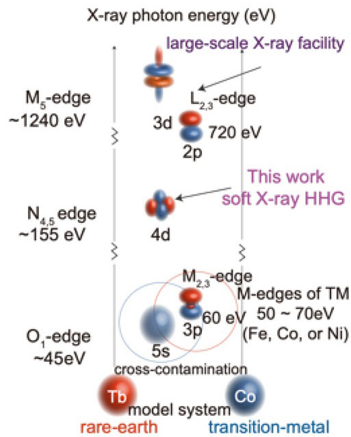
Illustration of the X-ray energies of different transition metals (TM) and rare-earth (RE) elements. [Image: G. Fan et al. “Ultrafast magnetic scattering on ferrimagnets enabled by a bright Yb-based soft x-ray source,” Optica 9, 399 (2022), doi: 10.1364/OPTICA.443440 (CC BY 4.0)] [Enlarge image]
The researchers used their laser system to analyze the behavior of a thin film of cobalt-terbium, targeting the 4d-absoprtion edge of terbium at about 155 eV. They did this by exciting the sample with infrared pulses at 1550 nm and using a CCD camera to record the diffraction patterns produced after irradiating the excited material with the X-rays. By recording a series of separate patterns one after the other, they were able to track the evolution of the material’s magnetic domains in space and time.
The researchers found that the material demagnetizes on two distinct timescales. About 10% of the sample loses it magnetization less than a picosecond after being excited by the infrared laser, while the rest of the domains become randomized over the course of several tens of picoseconds. They say that these results are in good agreement with data from a number of rare-earth material systems studied previously.
Future applications
“Before our work,” the researchers write, “similar experiments could be carried out only at X-ray free-electron lasers and femtosecond-slicing facilities.”
With this demonstration under their belt, the researchers say that their laser system could now be used to image and manipulate electron spin in a wider range of magnetic nanostructures, and, in particular, aid in the development of skyrmion devices. More broadly, they reckon that ytterbium lasers with ever higher average powers could be used in “photon-hungry” applications across the sciences. One possible application, they say, is to probe the electronic motion of biological molecules in liquids—their natural habitat.