
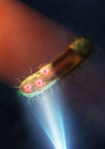
This illustration represents a bacterium being illuminated with mid-infrared in the top left, while visible light from a microscope underneath is used to help capture the image. [Image: 2024 Ideguchi et al./ Nature Photonics]
While mid-infrared (MIR) microscopy can provide label-free and nondestructive chemical analysis, one major drawback is its low spatial resolution of a few micrometers imposed by the diffraction limit. Recent advances in super-resolution MIR imaging, including MIR photothermal imaging, have allowed for the visualization of submicrometer structures of living cells and pathology slides.
Now, researchers from the University of Tokyo, Japan, have demonstrated far-field MIR spectroscopic imaging with a spatial resolution as low as 120 nm, which represents a 30-fold improvement on the resolution of typical MIR microscopes (Nat. Photon., doi: 10.1038/s41566-024-01423-0). Their approach—an implementation of wide-field MIR photothermal imaging—has the potential to cross over into the nanoscopic range, defined as a spatial resolution of less than 100 nm.
The trouble with MIR microscopy
Study author Takuro Ideguchi and his colleagues aimed to develop a label-free chemical nanoscopy technique that could measure living biological specimens without damage or toxicity. In particular, their main target is bacteria, which are micrometers in size. Imaging the structures inside living bacteria has wide-ranging applications, such as diagnosing infectious diseases, testing antimicrobial resistance and monitoring the production of biofuels.
“Mid-infrared microscopy is one of the vibrational microscopy techniques with which we can acquire image contrasts based on molecular vibrations,” said Ideguchi. “Since there is no need to use fluorescence probes or staining, it allows label-free chemical imaging.”
However, the long-wavelength MIR diffraction limit leads to an intrinsically low spatial resolution of several micrometers. MIR photothermal microscopy boosts the spatial resolution by combining MIR absorption and visible imaging. The absorption of MIR light due to molecular vibration leads to local heat, resulting in a change in the refractive index.
A visible-light microscope is then employed to visualize the local refractive index change with a resolution determined by the visible diffraction limit—at least in theory. In practice, typical MIR photothermal microscopes have spatial resolutions of several hundred nanometers, due to heat diffusion from the photothermal effect.
Their microscope, based on MIR photothermal quantitative phase imaging (MIP-QPI), implements a synthetic aperture technique in the QPI system that pushes the limit of spatial resolution.
Pushing the limit
Instead of a visible-light microscope, Ideguchi and his colleagues used the quantitative phase imaging (QPI) technique, a label-free approach that measures the optical phase delay of transparent specimens. Their microscope, based on MIR photothermal quantitative phase imaging (MIP-QPI), implements a synthetic aperture technique in the QPI system that pushes the limit of spatial resolution.
“Here, we take multiple QPI images with tilted illumination with different angles, which allows us to acquire larger spatial frequency information. We reconstruct an image with a higher spatial resolution by computing the acquired several images,” said Ideguchi. “With this scheme, we have achieved around 120-nm spatial resolution.”
The researchers also incorporated new MIR and visible-light laser systems with pulse durations of less than 1 ns, which reduces heat diffusion caused by the photothermal effect. Lastly, they tested the MIP-QPI microscope on two bacterial species, Escherichia coli and Rhodococcus jostii RHA1.
“Since our microscope’s spatial resolution is high enough, we can clearly visualize intracellular structures in bacteria with chemical contrast,” he said. “We have estimated that the spatial resolution of the microscope could be even improved down to less than 100 nm. It is also possible to make the imaging faster, potentially up to the video rate.”